Caries detection in the 21st century -- sharpening our diagnostic abilities

By Martin Jablow, DMD“A sharp explorer should be used with some pressure and if a very slight pull is required to remove it, the pit should be marked for restoration even if there are no signs of decay.” This is a quote from G.V. Black in 1924.1Caries is not as simple to diagnose today as it was decades ago when there were large, bombed out teeth. With the increased use of fluoride, the detection of caries is not as simple as it used to be. Yet in 2009 many of us are still diagnosing caries the same way as G.V. Black did in the early 1900s. The goal now is to be minimally invasive — catch caries at its earliest stages and attempt to remineralize incipient caries in teeth through the use of ozone and MI paste.Decay is difficult to detect in radiographs unless larger than 2 mm to 3 mm deep into dentin, or one-third the bucco-lingual distance.2 An explorer has high specificity for caries but low sensitivity for the caries. This means a lot of incipient caries can be missed if we rely on an explorer and radiographs alone.There is a call in the literature for discontinuance of the use of the dental explorer for caries detection. Some dental schools now teach reduced reliance on the explorer. An explorer may actually cause more harm by breaking the enamel rods when forced into an incipient carious lesion. At this early stage of caries remineralization should be considered. The problem is detecting the initial stage of caries.So what improved ways exist for the detection of caries? There are a number of different modalities for detecting caries. The most popular devices detect caries through the use of fluorescence. Normal healthy tooth structure produces little or no fluorescence. Carious tooth structure will fluoresce proportionate to the degree of caries. These devices are highly sensitive to caries but have a low specificity. This low specificity means that these devices will measure the fluorescence of anything. Caries detection devices are another tool aiding in the diagnosis of caries along with conventional diagnostic tools and good professional judgment. They can be used to monitor the progression of caries and aid in the decision to prevent, remineralize, or restore. The most popular device is the KaVo DIAGNOdent.
The DIAGNOdent is a 655 nm laser (the same as a red laser pointer) that detects fluorescence of decay in teeth. The DIAGNOdent will produce a value that can assist in the diagnosis of caries.
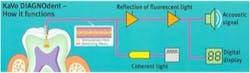
The DIAGNOdent emits an increasing audio tone and digital readout indicating the amount of caries present. The numbers can be recorded for a real world “watch” or to determine if remineralization is taking place. The DIAGNOdent is approved for use on smooth surface and pits and fissures only. It is not approved for interproximal use.A similar device is the Midwest Caries I.D. Instead of a laser it uses a light emitting diode (LED) to measure the caries reflection signature. The major difference is instead of a numerical readout, the Caries I.D. has a red and green indicator light for caries. This makes monitoring any progression of caries or remineralization more difficult. The device also beeps; the faster the beep, the more decay present. The Caries I.D. is approved for smooth surfaces, pits and fissures, and interproximal surfaces.Another device using florescence is Spectra from Air Techniques. Spectra uses a 405 nm LED, which causes porphyrins from caries-producing bacteria to fluoresce. Caries will produce a red color, and healthy teeth will fluoresce green. Spectra then produces a graphic and numerical display. The benefit of this device is that the graphic can be saved to imaging software, enhancing the ability to monitor caries and remineralization.

SOPROLIFE from Acteon enhances your ability to find caries by combining an intraoral camera for normal enhanced viewing through magnification and adds fluorescence technology to find caries. This device uses 450 nm light to cause the fluorescence. The device has both a diagnosis mode and a treatment mode. Diagnosis mode allows magnification from 30 to 100 times using white light. Turning a switch on the handpiece changes the lighting to diagnosis mode. This allows you to view healthy tooth structure vs. caries. Caries shows up red.

You can use this mode during caries excavation to determine if all the caries has been removed. At the time of this writing, 510K Clearance is pending on SOPROLIFE. Please contact Acteon to check FDA Clearance and item availability.
Another promising technology is the Canary Dental Caries Detection System. It uses a low power laser to scan the tooth for decay. The tooth absorbs the laser light and two phenomena are observed: the laser light is converted into luminescence, and there is a release of heat (less than 1 degree Celsius). This heat will not harm the tooth but gives important information on the tooth up to a depth of 5 mm below the surface. Simultaneous measurement of the reflected heat and light provides us with information on the presence and extent of tooth decay below the tooth surface.Another caries detection device on the horizon is Lantis Laser’s Optical Coherence Tomography (OCT) Dental Imaging System. OCT devices are capable of imaging both the teeth and periodontium. Using the hand-held scanner, the operator captures cross-sectional images, or tomographic slices, at up to 3 mm deep into dental tissue. These cross-sectional images are then displayed individually in real time on a chairside monitor and can be saved to the patient digital file.
The OCT device is capable of detecting recurrent decay around restorations, along with examining the marginal integrity of restorations bonded to tooth structure.As you can see, there are adjuncts to the current methods of caries detection. New methods will be appearing shortly that will improve the dentist’s ability to detect caries earlier than ever before. With this enhanced knowledge, we will be able to establish better protocols for caries intervention and treatment.
Martin Jablow, DMD, received his dental degree from New Jersey Dental School in 1986 and practices in Woodbridge, N.J. He received his fellowship in the Academy of General Dentistry and is certified in various laser wavelengths. He is a member of the American Dental Association and the New Jersey Dental Association. He is an attending dentist at John F. Kennedy Medical Center in Edison, N.J., as well as a long-time member of his county’s Peer Review Committee. Dr. Jablow is president of Dental Technology Solutions, a lecture and consulting company. Dr. Jablow is the writer of the Dr. Bicuspid.com Question and Answer column called “Ask Marty.” He also writes a monthly column for Dental Learning Hub’s Apex magazine. Dr. Jablow has spoken at many major dental meetings including the ADA Annual Session, the Yankee Dental Congress, and the California Dental Association. Consulting with various high-technology companies, Dr. Jablow is involved in the process of improving established products and testing new products and techniques. He consults with dental offices on implementing computers and technology into their practices. Dr. Jablow enjoys promoting the use of technology in the dental office through lecturing and writing articles for various national dental publications, with an emphasis on improving patient care.
References1 Black GV. Operative Dentistry. 1924; 1(7) 32.2 Rock WP, Kidd EAM. Br Dent J. 1988; 164(8) 243-247.
References1 Black GV. Operative Dentistry. 1924; 1(7) 32.2 Rock WP, Kidd EAM. Br Dent J. 1988; 164(8) 243-247.