Cone beam imaging is radically transforming dentistry
By Eugene Antenucci, DDSIn 1999 Bill Gates wrote a prophetic book titled “Business @ The Speed of Thought: Using A Digital Nervous System,” where he stated in the very first chapter: “I have a simple but strong belief. The most meaningful way to differentiate your company from your competition, the best way to put distance between you and the crowd, is to do an outstanding job with information. How you gather, manage and use information will determine whether you win or lose.”The relentless onward march of advanced digital technology in dentistry is a fact of life in managing a dental practice, and it is undeniably true that how we “gather, manage and use information will determine whether you win or lose.” (Gates, William H. III; Business @ The Speed of Thought: Using a Digital Nervous System, 1999; Warner Books, Inc.; p 4.)Cone beam imaging, while a relatively new technology to dentistry, has already radically transformed the way that we as dentists can gather information, with the result being an ability to diagnose and treatment plan in a manner that previously could not even be imagined. The transformation from interpreting two-dimensional information to diagnosing from 3-D imaging, which allows for visualization of all structures in any given field, is a quantum leap ... and one that has forever altered the way we must practice dentistry.Cone Beam Volumetric Tomography is a diagnostic imaging technology that uses radiation in a manner similar to conventional radiographic imaging, with the difference being that cone beam images are converted into a three-dimensional view that can then be manipulated by sophisticated computer software for a wide variety of applications, including implant, orthodontic, orthognathic TMJ, and diagnostic purposes.Cone beam imaging evolved from the work by Godfrey Hounsfield in the 1970s with the development of CAT Scan imaging in medicine. CAT Scans, or Computerized Axial Tomography, utilize a fan-shaped beam of radiation that rotates around the patient hundreds of times per second, with the patient being moved a precise distance in the scanner. Cone beam differs in that cone beam scans use a cone-shaped beam that is directed at a detector that rotates around the patient. Unlike CAT scans, these can be applied to dental practices.The units themselves can be similar in size to dental panoramic and cephalometric units. Cone beam units are within the range of affordability for dental practices. Patient radiation exposure is significantly lower, as is the cost per image to patients. The technology is relatively easy to implement and use, and accuracy and precision is high with a 1:1 display of dental structures. The technology is easy to learn, and the software applications accessible for dentistry are extensive. The end result for dentistry is an affordable means of elevating the level of diagnostic abilities within the dental practice. With cone beam-produced images, hundreds of “slices” of information are gathered and assembled by software into three-dimensional views of any aspect of any given study. Data can be imported into software, such as Romexis by Planmeca, for the purpose of comprehensive diagnostic analysis and presurgical treatment planning, as well as for the production of stereolithographic biomodels. Cone beam images are reconstructed by software into three-dimensional planes, each of which allow for interaction and manipulation. The diagnostician has the ability to specify precisely which section of the study will be visualized in each plane.
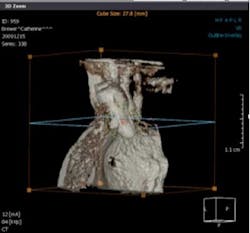
Images of coronal, axial, and sagittal planes, as well as enhanced VR imageIn 2008, I incorporated the Planmeca ProMax 3D into our office. My practice, Huntington Bay Dental, is a general dental practice that provides a full range of restorative and surgical services to patients of all ages. Along with my partner and associate dentists, we routinely place as well as restore dental implants.Our decision to introduce cone beam technology was initially based on the knowledge that the state-of-the-art in implant treatment planning involved the use of three-dimensional imaging; however, our patients were forced to go to outside facilities for this service. CAT scans at imaging centers provided us with a great deal of information, but at a greater cost to the patient in terms of both higher radiation and more dollars.We selected the Planmeca ProMax 3D because of its smaller footprint size, its accuracy, and its ability to take both a two-dimensional panorex as well as a three-dimensional cone beam image. As an instrument for treatment planning implant cases, it exceeded all of our expectations, with the added bonus being that we were able to take more cases from diagnosis to completion — patients not only saved money, but they also did not get “lost” in the referral process for imaging. To our surprise, however, we found that 3-D imaging became indispensible in a wide range of dental diagnosis and treatment planning, to the point that there are many cases today where we cannot even imagine understanding how to best help a patient without the use of the third dimension. We found applications in endodontics with visualizing fractured roots, periapical pathology, and accessory canals; in treatment planning for the extraction of third molars in proximity to the mandibular nerve; in diagnosing pathology of all types; in understanding the cause of previously undiagnosed pain; in visualizing sinus pathology; in understanding the etiology of temporomandibular joint dysfunctions; and in diagnosing the true extent of periodontal pathology and disease in localized areas. And while we do not practice orthodontics, we are well aware of the orthodontic applications of 3-D imaging.In implant treatment planning, we have come to hold the strong opinion that the standard of care today in implant dentistry is to recommend 3-D imaging for implants for nearly every patient, and because of this it would be poor practice to do implants without using 3-D imaging. This philosophy has quickly expanded into all phases of our practice of dentistry. I am by no means implying that 3-D images are required for all patients, but rather am stating that I strongly believe that there are cases where two dimensions are simply not enough, and the addition of the third dimension — when applicable — becomes the only means of providing an accurate diagnosis. The presence of this technology has forever altered the standard of care in our profession with respect to diagnosis and treatment planning. 3-D cone beam imaging has raised the bar and redefined the standard of care for many areas of dental practice. The World War I song from 1919 by Byron G. Harlan with Ada Jones titled “How Ya Gonna Keep 'Em Down On the Farm After They've Seen Paree” just about sums up my view regarding the presence of cone beam imaging in dentistry. The technology is here in an affordable and ergonomic form, providing diagnostic capabilities far beyond what is available with 2-D. When indicated, there simply is no sound excuse for not incorporating the technology. And the words written by Bill Gates in 1999 ring true: “How you gather, manage and use information will determine whether you win or lose.”For dentistry, “winning and losing” is not primarily a financial consideration. Winning and losing ultimately deals with the quality of care that we provide our patients, and doing all within our means to assure the best outcome for every procedure we perform. The third dimension in dentistry raises the odds for consistent success.Does visualization of the third dimension really make a difference?

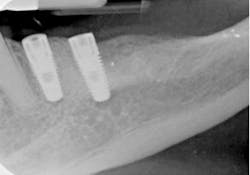
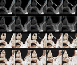
These two-dimensional digital periapical images were taken intraoperatively during implant placement. They show two implants positioned parallel to each other with no apparent problems with their surgical placement.
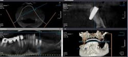
Cone-beam imaging postoperatively illustrate the importance of the third dimension by clearly showing that two-dimensional images show only a portion of the imaged site’s reality. Although parallel from a frontal view in 2-D, the mesial implant is clearly angled from a buccal-lingual perspective and has perforated the lingual plate of bone.

In implant treatment planning, even for simple cases, cone beam imaging allows for concise and predictable treatment planning. Anatomic landmarks such as foramina and the mandibular canal can be precisely marked, and cross-sectional views accurately show the width and length of available bone, give indications to the thickness and density of the bone, and provide virtual implant placement of virtually all implants from software libraries.
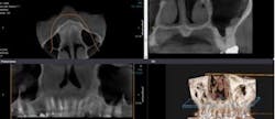
This scan was taken to evaluate the #7 site for placement of an implant due to the fracture of the tooth. The scan reveals the presence of a mucous retention cyst in the left sinus space.
Sectional views of the case clearly show the location and size of the cyst on the walls of the sinus.
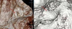
Software tools can be used to “enter” the sinus itself and clearly visualize the lesion virtually.

Eugene Antenucci, DDS, is a general dentist who maintains a full-time private practice in Huntington, N.Y. His state-of-the-art dental facility is also home to a continuing dental education training center for dentists, as well a commercial dental laboratory. His practice, Huntington Bay Dental, was distinguished as “Dental Practice of the Month” by Dental Economics in May 2003 and “Business of the Year” by the Huntington Chamber of Commerce for 2003. Dr. Antenucci is a 1983 graduate of New York University College of Dentistry. He was awarded his Fellowship in the Academy of General Dentistry in 1992, the American College of Dentists in 1999, and the International College of Dentists in 2005. Dr. Antenucci is a certified CEREC Basic and Advanced Training Instructor, and has conducted training seminars throughout the United States. He lectures internationally, conducting seminars in the clinical utilization of advanced technology in dentistry, as well as seminars in cosmetic dentistry, practice management, CEREC, and laser training. Dr. Antenucci serves on the Board of Benefactors of the Guide Dog Foundation and America’s Vet Dogs, and is also an active member of the National Italian American Foundation, serving as the New York Area Coordinator for the organization. Dental equipment manufacturer Planmeca USA has retained Dr. Antenucci as a spokesperson for its line of 3-D imaging products and to advise the company on marketing, advertising, and continuing-education efforts.