DIAGNOdent caries detection aid
By Maria Perno Goldie, RDH, MS
While 80% of tooth decay lesions occurs on the occlusal surface of the tooth, a considerable percentage of these lesions go undetected using conventional procedures and instruments. Conventional methods of diagnosing dental decay, such as use of the dental explorer or bite-wing radiographs, are often ineffective in detecting enamel defects, as they may be too small or inaccessible to the diagnostic tool.(1) Observations show sensitivity and specificity values of 62 and 84 percent with the explorer and visual examination, respectively, indicating that clinicians were more likely to fail to treat carious fissures than to restore sound fissures.(1) A number of diagnostic decisions to place occlusal restorations are based on the inappropriate use of the dental explorer to determine the softness or tackiness of the fissure or the amount of resistance to the removal of the explorer from the fissure.(2) The conclusion of one study, and corroborating evidence by other studies, state that there is strong evidence to support the elimination of the use of the dental explorer as the primary diagnostic tool in diagnosing dental decay. However, the explorer can still be a valuable tool in removing debris, feeling the margins of existing restorations, and other tactile tasks.We now know that the caries process is a continuum and if detected before cavitation is noted, the process may be able to be reversed or arrested with a number of products and treatments. If our goal is prevention, the nature of the clinical examination for dental decay has changed from the detection of advanced tooth decay requiring restoration, to the identification of lesions or demineralized areas at the precavitation stage that may be reversed or arrested if the thin surface layer covering the demineralized area remains intact.(3) Thus, the use of the dental explorer in the traditional manner must be avoided, because it will fracture the surface layer and eliminate the possibility of reversing the caries process.(3)
While 80% of tooth decay lesions occurs on the occlusal surface of the tooth, a considerable percentage of these lesions go undetected using conventional procedures and instruments. Conventional methods of diagnosing dental decay, such as use of the dental explorer or bite-wing radiographs, are often ineffective in detecting enamel defects, as they may be too small or inaccessible to the diagnostic tool.(1) Observations show sensitivity and specificity values of 62 and 84 percent with the explorer and visual examination, respectively, indicating that clinicians were more likely to fail to treat carious fissures than to restore sound fissures.(1) A number of diagnostic decisions to place occlusal restorations are based on the inappropriate use of the dental explorer to determine the softness or tackiness of the fissure or the amount of resistance to the removal of the explorer from the fissure.(2) The conclusion of one study, and corroborating evidence by other studies, state that there is strong evidence to support the elimination of the use of the dental explorer as the primary diagnostic tool in diagnosing dental decay. However, the explorer can still be a valuable tool in removing debris, feeling the margins of existing restorations, and other tactile tasks.We now know that the caries process is a continuum and if detected before cavitation is noted, the process may be able to be reversed or arrested with a number of products and treatments. If our goal is prevention, the nature of the clinical examination for dental decay has changed from the detection of advanced tooth decay requiring restoration, to the identification of lesions or demineralized areas at the precavitation stage that may be reversed or arrested if the thin surface layer covering the demineralized area remains intact.(3) Thus, the use of the dental explorer in the traditional manner must be avoided, because it will fracture the surface layer and eliminate the possibility of reversing the caries process.(3)
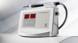
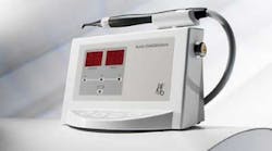
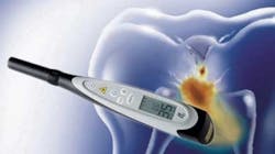
With the introduction of new detection devices, such as the DIAGNOdent, along with the use of traditional instruments and treatments, clinicians can more effectively identify the presence of occlusal decay and properly treat the tooth structure, possible in a non-invasive manner. The technology of the DIAGNOdent, a 655 nm diode laser, allows for detection of noncavitated, occlusal pit-and-fissure tooth decay, in addition to smooth surface caries at an earlier stage than visual inspection.(4) The DIAGNOdent measures laser fluorescence within the mineral structure of the tooth. As the incident laser light is disseminated into the site, two-way handpiece optics allows the unit to simultaneously quantify the reflected laser light energy.(5) At the specific wavelength that the DIAGNOdent laser operates, healthy tooth structure exhibits little or no fluorescence, resulting in very low scale readings on the display. However, decayed tooth structure will exhibit fluorescence, proportionate to the degree of lost tooth structure, resulting in elevated scale readings on the display of the DIAGNOdent. An audio signal allows the operator to hear changes in the scale values, enabling focus on the patient and not solely on the device.(5) This is beneficial not only for detection of decay, but also for patient acceptance of treatment plans.Recommendations for treatment are: Values between 10–15 require no active care or treatment; Values between 15–30 require preventative or operative care, depending on the patient’s caries risk; Values of 30+ require operative and preventative care.(5)
One study compared the accuracy and repeatability of three diagnostic systems (DIAGNOdent, visual and radiographic) for occlusal caries diagnosis in primary molars.(6) The DIAGNOdent was the most accurate system in the study for the detection of occlusal dentinal decay in primary molars. The performance of the DIAGNOdent systems was not statistically significantly better than that achieved using visual examination for non-cavitated teeth. The conclusion was that the DIAGNOdent may prove useful as a predictive clinical tool. The authors also noted that, with appropriate training, visual examination may offer similar results without the need for additional equipment.(6)Analysis of available data in 2004 suggested that DIAGNOdent is a reproducible method for caries detection, with good sensitivity and specificity.(7) It is most suitable at the advanced stages of D2 and D3 levels, and is most appropriate for tooth decay detection on occlusal and accessible smooth surfaces. Distinct fluorescence of the tooth decay in more advanced stages, D2, D3, leads to the assumption that, besides light scattering, bacteria or their metabolites could contribute to the fluorescence of these lesions. This hypothesis was tested and validated.(8) A follow-up study results also suggested that DIAGNOdent is useful in monitoring occlusal caries in both permanent and primary molars.(9) In conclusion, the DIAGNOdent is a valuable adjunct to clinical examination. References
1. Lussi A. Validity of diagnostic and treatment decisions of fissure caries. Caries Res 1991; 25:296-303.2. Anusavice K. The maze of treatment decisions. In: Fejerskov O, Kidd EA, eds. Dental caries. The disease and its clinical management. Malden, Mass.: Blackwell; 2003:251-65.3. Hamilton JC and Stookey G. Should a dental explorer be used to probe suspected carious lesions? J Am Dent Assoc 2005; 136; 1526-1532.4. Lussi A. Clinical performance of the laser fluorescence system DIAGNOdent for detection of occlusal caries (in German). Acta Med Dent Helv 2000; 5:15-19.5. Sanchez-Figueras A. Laser Fluorescence Detection of Occlusal Caries. Clinical utilization of the KaVo DIAGNOdent. www.kavo.com/img_cpm/global/files/009/diagnodent/DIAGNOdent_LaserDetection.pdf.6. Attrill DC, Ashley PF. Occlusal caries detection in primary teeth: a comparison of DIAGNOdent with conventional methods. Br Dent J. 2001 Apr 28; 190(8):440-3.7. Lussi A, Hibst R, Paulus R. DIAGNOdent: An Optical Method for Caries Detection. JDR July 2004 vol. 83 no. suppl 1 C80-C83.8. Hibst R, Paulus R, Lussi A (2001). Detection of occlusal caries by laser fluorescence. Basic and clinical investigations. Med Laser Appl 16:205–213.9. Anttonen, V., Seppä, L. and Hausen, H. (2004), A follow-up study of the use of DIAGNOdent for monitoring fissure caries in children. Community Dentistry and Oral Epidemiology, 32: 312–318. More recent studies: www.kavousa.com/Default.aspx
1. Lussi A. Validity of diagnostic and treatment decisions of fissure caries. Caries Res 1991; 25:296-303.2. Anusavice K. The maze of treatment decisions. In: Fejerskov O, Kidd EA, eds. Dental caries. The disease and its clinical management. Malden, Mass.: Blackwell; 2003:251-65.3. Hamilton JC and Stookey G. Should a dental explorer be used to probe suspected carious lesions? J Am Dent Assoc 2005; 136; 1526-1532.4. Lussi A. Clinical performance of the laser fluorescence system DIAGNOdent for detection of occlusal caries (in German). Acta Med Dent Helv 2000; 5:15-19.5. Sanchez-Figueras A. Laser Fluorescence Detection of Occlusal Caries. Clinical utilization of the KaVo DIAGNOdent. www.kavo.com/img_cpm/global/files/009/diagnodent/DIAGNOdent_LaserDetection.pdf.6. Attrill DC, Ashley PF. Occlusal caries detection in primary teeth: a comparison of DIAGNOdent with conventional methods. Br Dent J. 2001 Apr 28; 190(8):440-3.7. Lussi A, Hibst R, Paulus R. DIAGNOdent: An Optical Method for Caries Detection. JDR July 2004 vol. 83 no. suppl 1 C80-C83.8. Hibst R, Paulus R, Lussi A (2001). Detection of occlusal caries by laser fluorescence. Basic and clinical investigations. Med Laser Appl 16:205–213.9. Anttonen, V., Seppä, L. and Hausen, H. (2004), A follow-up study of the use of DIAGNOdent for monitoring fissure caries in children. Community Dentistry and Oral Epidemiology, 32: 312–318. More recent studies: www.kavousa.com/Default.aspx

Maria Perno Goldie, RDH, MS