The Canary System: a solution for detecting caries around restoration margins

By Dr. Stephen Abrams
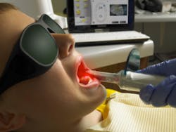
Figure 1 The Canary System
Caries is one of the major oral diseases treated in clinical practice. It not only destroys tooth structure but can undermine restorations. Detecting caries around restoration margins is challenging if not impossible as the materials impede or block all forms of energy.
Caries is one of the major oral diseases treated in clinical practice. It not only destroys tooth structure but can undermine restorations. Detecting caries around restoration margins is challenging if not impossible as the materials impede or block all forms of energy.
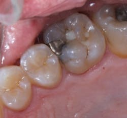
Figure 2 Occlusal view of Maxillary Right First Molar showing the amalgam and composite restorations. The margins appear intact around the amalgam The Canary System, using PTR-LUM energy conversion technology, is able to examine the restoration margin and detect caries along this interface earlier than would be detected with radiographs. Rapid, safe pulses of laser light combining infrared and luminescence allow one to examine lesions as small as 50 microns and subsurface caries up to 5 mm below the tooth surface or along the margin of composites and amalgams.

Figure 3 Bitewing Radiograph taken 4 weeks before the restoration was replaced. No sign of caries beneath the restorations on the maxillary first molar At our recall examination, we were concerned about the grey colouration around the amalgam on the mesial surface of the maxillary first molar. A bitewing radiograph did not reveal any sign of caries around the amalgam restoration and composite resin restoration in the mesial pit was not well delineated in the radiograph. A Canary Scan of the amalgam indicated caries along the buccal and distal margins of the restoration.

Figure 4 Canary Scan done at the time of the bitewing radiograph.
Upon removal of the amalgam there was a large area of caries along the buccal margin, distal margin and extending on to the gingival floor of the preparation. The buccal caries had begun to undermine the mesio-buccal cusp. If this had not been detected, the patient, at a minimum, would have fractured the buccal cusp.
Upon removal of the amalgam there was a large area of caries along the buccal margin, distal margin and extending on to the gingival floor of the preparation. The buccal caries had begun to undermine the mesio-buccal cusp. If this had not been detected, the patient, at a minimum, would have fractured the buccal cusp.
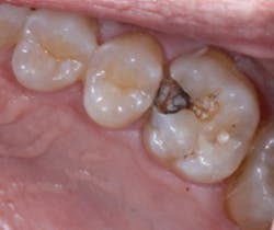
Figure 5 The amalgam restoration removed showing caries along the gingival seat and the buccal and lingual walls of the preparation
Over ten years of research on PTR-LUM technology and two clinical trials have demonstrated that The Canary System can detect caries in the following situations:
• Occlusal Pit and fissure caries(1,2,3)
• Smooth Surface Caries(4,5)
• Acid Erosion Lesions(6,7)
• Root Caries(8,9)
• Interproximal caries lesions(10,11)
• Demineralization and Remineralization of early caries lesions(12,13,14,15)
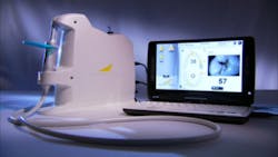
Figure 6 The Canary System
The Canary System captures both signals and images of the tooth surfaces being examined. These images are displayed on an interactive, touch-screen monitor for immediate chair side review with the patient who can also take home a printed Canary report. The Canary System creates an opportunity for dialogue and co-diagnosis with the patient, two strong motivators for a new long-term, recurring patient relationship. Odontograms also are added to patients’ files so treatment progress can be monitored over time. The data is saved on the computer, and merged into detailed user-friendly reports.
The Canary System captures both signals and images of the tooth surfaces being examined. These images are displayed on an interactive, touch-screen monitor for immediate chair side review with the patient who can also take home a printed Canary report. The Canary System creates an opportunity for dialogue and co-diagnosis with the patient, two strong motivators for a new long-term, recurring patient relationship. Odontograms also are added to patients’ files so treatment progress can be monitored over time. The data is saved on the computer, and merged into detailed user-friendly reports.

Figure 7 The Ideal Characteristics of a Caries Detection System
The essential characteristics of the ideal caries detection system are: 1. High sensitivity and specificity for caries detection. Existing detection systems often demonstrate an inverse relationship between sensitivity and specificity. Where radiographs suffer from sensitivity they make up in specificity.(16) Fluorescence-based systems, such as DIAGNOdent, have high sensitivities, however have poor specificity. A device that can balance and provide both high sensitivity and specificity are ideal for a caries detection system to limit false positive and false negative diagnoses. 2. The ability to detect and monitor both de- and remineralization of carious lesions. Detection of small early carious lesions that are at a minimum a few hundred microns in size up to a few millimetres below the tooth surface may be difficult to detect or monitor especially if they are surrounded by intact tooth structure. One also needs to consider how oral bacteria and surface stain influence the measurements of the particular detection technology. 3. Ability to detect lesions on all surfaces including smooth surfaces, root surfaces, occlusal surfaces and interproximal regions. Having the ability to detect pit and fissure caries while neglecting smooth surface or interproximal areas really limits the clinician to detecting only a small portion of the potential disease sites. 4. Ability to detect caries around restorations. In older patient groups this is where a large number of caries occur. Detection around the visible margins is one area. Detection of caries at the gingival seat of Class II restoration or along the walls of the proximal box is very challenging. Radiographs do detect caries along the gingival seat but only when a large lesion is present. The challenge in both situations is the presence of a highly reflective material such as amalgam or composite resin that may complicate detection systems by limiting light penetration and detection of subsurface decay. . 5. Non-invasive & non-harmful technology is essential particularly for those employing remineralization programs. With The Canary System, a new infrared and fluorescence based system, one can detect changes in lesion size at one month after starting remineralization.(17) 6. Repeatable measurements; measurements that are linked to the lesion size or crystal structure. This is a key characteristic since one is detecting and treating changes in enamel crystal structure not detecting the presence of oral bacteria trapped below the tooth surface that may or may not be carious-causing bacteria, such as lactobacillus and strep mutans strains. 7. Imaging or image capture that will allow the operator to see the surface under examination. 8. System for recording measurements and monitoring changes in measurements over time. Ideally, the measurement recording should be contained within the device to allow for easy retrieval of patient history. This is essential for recordkeeping as well. Furthermore, for those devices with independent software, integration with all practice management programs is a necessity for facilitating integration into clinical practice. 9. A system that will help motivate patients to improve their oral health care and provide them with tools and information. Engaging and empowering patients is the key to the success of any preventive based program. 10. A system based on extensive in vitro investigations and clinical data published in peer-reviewed journals, including FDA/ Health Canada approved clinical trials demonstrating the safety and effectiveness of the system to detect and monitor carious lesions. 11. Minimal or no preparation of the tooth surface prior to taking a reading. Cleaning, polishing and creating a dry field before measuring a tooth surface all add time and cost to any diagnostic procedure. Finding a device that can work in the oral environment with minimal tooth surface preparation translates into efficient use of operator time. 12. Ability to detect and monitor erosion lesions. Although these are not true carious lesions and are formed in a much different fashion, a system that monitors both caries and erosion will provide the clinician with a wide range of conditions in clinical practice. A device with these twelve characteristics should provide the user with a very robust method for caries detection. The ability to detect small carious lesions and monitor changes will allow the provider to shift the emphasis from placement of restorations to remineralization of early carious lesions.
The essential characteristics of the ideal caries detection system are: 1. High sensitivity and specificity for caries detection. Existing detection systems often demonstrate an inverse relationship between sensitivity and specificity. Where radiographs suffer from sensitivity they make up in specificity.(16) Fluorescence-based systems, such as DIAGNOdent, have high sensitivities, however have poor specificity. A device that can balance and provide both high sensitivity and specificity are ideal for a caries detection system to limit false positive and false negative diagnoses. 2. The ability to detect and monitor both de- and remineralization of carious lesions. Detection of small early carious lesions that are at a minimum a few hundred microns in size up to a few millimetres below the tooth surface may be difficult to detect or monitor especially if they are surrounded by intact tooth structure. One also needs to consider how oral bacteria and surface stain influence the measurements of the particular detection technology. 3. Ability to detect lesions on all surfaces including smooth surfaces, root surfaces, occlusal surfaces and interproximal regions. Having the ability to detect pit and fissure caries while neglecting smooth surface or interproximal areas really limits the clinician to detecting only a small portion of the potential disease sites. 4. Ability to detect caries around restorations. In older patient groups this is where a large number of caries occur. Detection around the visible margins is one area. Detection of caries at the gingival seat of Class II restoration or along the walls of the proximal box is very challenging. Radiographs do detect caries along the gingival seat but only when a large lesion is present. The challenge in both situations is the presence of a highly reflective material such as amalgam or composite resin that may complicate detection systems by limiting light penetration and detection of subsurface decay. . 5. Non-invasive & non-harmful technology is essential particularly for those employing remineralization programs. With The Canary System, a new infrared and fluorescence based system, one can detect changes in lesion size at one month after starting remineralization.(17) 6. Repeatable measurements; measurements that are linked to the lesion size or crystal structure. This is a key characteristic since one is detecting and treating changes in enamel crystal structure not detecting the presence of oral bacteria trapped below the tooth surface that may or may not be carious-causing bacteria, such as lactobacillus and strep mutans strains. 7. Imaging or image capture that will allow the operator to see the surface under examination. 8. System for recording measurements and monitoring changes in measurements over time. Ideally, the measurement recording should be contained within the device to allow for easy retrieval of patient history. This is essential for recordkeeping as well. Furthermore, for those devices with independent software, integration with all practice management programs is a necessity for facilitating integration into clinical practice. 9. A system that will help motivate patients to improve their oral health care and provide them with tools and information. Engaging and empowering patients is the key to the success of any preventive based program. 10. A system based on extensive in vitro investigations and clinical data published in peer-reviewed journals, including FDA/ Health Canada approved clinical trials demonstrating the safety and effectiveness of the system to detect and monitor carious lesions. 11. Minimal or no preparation of the tooth surface prior to taking a reading. Cleaning, polishing and creating a dry field before measuring a tooth surface all add time and cost to any diagnostic procedure. Finding a device that can work in the oral environment with minimal tooth surface preparation translates into efficient use of operator time. 12. Ability to detect and monitor erosion lesions. Although these are not true carious lesions and are formed in a much different fashion, a system that monitors both caries and erosion will provide the clinician with a wide range of conditions in clinical practice. A device with these twelve characteristics should provide the user with a very robust method for caries detection. The ability to detect small carious lesions and monitor changes will allow the provider to shift the emphasis from placement of restorations to remineralization of early carious lesions.

The unique characteristics of The Canary System provide a clinical practice with the ability to monitor the entire spectrum of the caries process; from initial demineralization to caries beneath pits and fissures and around restoration margins. The Canary System will enable a practice to provide preventive minimally invasive oral health care. Disclosure
Dr. Stephen Abrams is the CEO and Co-Founder of Quantum Dental Technologies which has developed The Canary System mentioned in this article. He has not received any compensation for the preparation of this article.References
1. Jeon, R. J., Han, C., Mandelis, A., Sanchez, V., Abrams, S. H., “Dental Depth Profilometric Diagnosis of Pit & Fissure Caries using Frequency-Domain Infrared Photothermal Radiometry and Modulated Laser Luminescence”, Early Detection of Caries III Proceedings of the Sixth Indiana Conference Indiana University School of Dentistry, pages 49 – 67, Stookey, G., editor, 2003 2. Jeon, R. J., Han, C., Mandelis, A., Sanchez, V., Abrams, S. H., “Diagnosis of Pit & Fissure Caries using Frequency-Domain Infrared Photothermal Radiometry and Modulated Laser Luminescence” Caries Research 2004; 38: 497- 513. 3. R. Jeon, A. Mandelis and S. Abrams, “Dental depth profilometric diagnosis of pit and fissure caries using frequency-domain infrared photothermal radiometry and modulated luminescence”, SPIE Vol. 5320, Photons plus Ultrasound: Imaging and Sensing (A. A. Oraevsky and L. V. Wang, Eds., Bellingham WA 2004, pp. 29 39 4. Jeon, R. J., Han, C., Mandelis, A., Sanchez, V., Abrams, S. H., “Non-intrusive, Non-contacting Frequency-Domain Photothermal Radiometry and Luminescence Depth Profilometry of Carious and Artificial Sub-surface Lesions in Human Teeth,” Journal of Biomedical Optics 2004, July – August ,9, # 4, 809 – 819. 5. Jeon, R. J., Mandelis, A., Abrams, S. H., “Depth profilometric case studies in caries diagnostics of human teeth using modulated laser radiometry and luminescence”, Review of Scientific Instruments, 2003; 74 (1): 380 - 383 6. Jeon R. J., Phan T. D. T., Wu A., Kulkarni G., Abrams S. H., and Mandelis A., “Photothermal radiometric quantitative detection of the different degrees of demineralization of dental enamel by acid etching,” J. Physique IV France, 2005; 125: 721 – 72 7. Abrams, SH., Matvienko, A., Ye, V., Mandelis, A., Ramalingam K., Amaechi, BT. "Detection and monitoring of dental erosion using PTR-LUM". International Association of Dental Research (IADR), Abstract # 238 [San Diego, CA, USA] March 2011 8. Jeon, R. J. Hellen, A., Matvienko, A., Mandelis, A., Abrams, S. H., Amaechi, B. T.., ”Detection of Demineralized-Remineralized Lesions on Root and Enamel of Human Teeth in vitro using Infrared Photothermal Radiometry and Modulated Luminescence”, ORCA Abstract # 157, Caries Research 2007, 41:323 9. Jeon R. J., Hellen A., Matvienko A., Mandelis A., Abrams S. H., Amaechi B. T., “In vitro Detection and Quantification of Enamel and Root Caries Using Infrared Photothermal Radiometry and Modulated Luminescence.” Journal of Biomedical Optics, 2008; 13(3), 048803, 10. Jeon R.J., Matvienko A., Mandelis A., Abrams S.H., Amaechi B.T, Kulkarni G. “Detection of interproximal demineralized lesions on human teeth in vitro using frequency-domain infrared photothermal radiometry and modulated luminescence”, J. BioMed. Optics, 2007; 12(3); 034028 1 – 13 11. Jeon, R. J., Matvienko, A., Mandelis, A., Abrams, S. H., Amaechi, B. T., Kulkarni, G., “Interproximal Dental Caries Detection using Photothermal Radiometry (PTR) and Modulated Luminescence (LUM)”, European Physical Journal, Special Topics, 2008, 153; 467 – 469 12. Jeon, R. J. Hellen, A., Matvienko, A., Mandelis, A., Abrams, S. H., Amaechi, B. T.., ”Detection of Demineralized-Remineralized Lesions on Root and Enamel of Human Teeth in vitro using Infrared Photothermal Radiometry and Modulated Luminescence”, ORCA Abstract # 157, Caries Research 2007, 41:323 13. Matvienko, A. Mandelis, A., Hellen, A., Jeon, R. J., Abrams S. H., Amaechi, B., “Quantitative Analysis of Incipient Mineral Loss in Hard Tissues”, (SPIE BiOS, San Jose, USA, January 2009), Proc. SPIE BiOS Vol. 7166 (12), 71660C1 – 12 (April 2009). 14. Hellen, A. Mandelis, A and Y. Finer, “Photothermal Radiometry and Modulated Luminescence Examination of Demineralized and Remineralized Lesions”, J Phys: Conf Ser 2010; 214(1): 012024 [15th International Conference on Photoacoustic and Photothermal Phenomena, Leuven, Belgium, July 2009] 15. Matvienko, A., Amaechi, BT., Ramalingam, K., Macaden, M., Ye, V., Hellen, A., Jeon, RJ., Sivagurunathan, K., Mandelis, A., Abrams, SH., "PTR-LUM-based detection of demineralization and remineralization of human teeth". International Association of Dental Research (IADR), Abstract # 114 [San Diego, CA, USA] March 2011. 16. Pretty, I. A., Maupome, G., “A Closer Look at Diagnosis in Clinical Dental Practice: Part 3. Effectiveness of Radiographic Diagnostic Procedures”, JCDA, 2004; 70(6): 388 – 394. 17. Sivagurunathan, K., Abrams, S. H., Garcia, J., Mandelis, A., Amaechi, B. T., Finer, Y., Hellen, W. M. P., and Elman, G.. “PTR-LUM (“The Canary System”) Clinical Trial Results for Caries Detection”. International Association of Dental Research, Caries Detection Session; Abstract # 3745 [Barcelona, Spain, July 14 - 17, 2010].
Stephen Abrams is a general dental practitioner with over 30 years of clinical experience. Dr. Abrams was awarded the Barnabus Day Award from the Ontario Dental Association for 20 years of distinguished service to the dental profession.