Lesion Ctg 1

Subgingival anomalies and pathologies viewed with the Perioscope (Part 2)

Dec. 21, 2012
In a second installment, John Kwan, DDS, and Suzanne Newkirk, RDH, continue the exploration of various anomalies and pathologies visualized with the Perioscope that have been found to be contributing factors in localized periodontal breakdown. Included are a pair of resorption case studies by Newkirk and Dr. Robert Gottlieb plus two items about the treatment of root resorption by Dr. Kwan.

Case #2: By Dr. Robert Gottlieb and Suzanne Newkirk, RDH

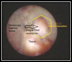
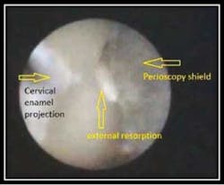
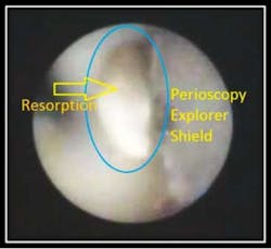
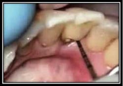
A patient with advanced periodontal disease, initially referred for a CTG (connective tissue graft) on the buccal of #2

Resorptive lesion found during initial debridement

Pre-Tx PC #2 Dec.9,2010

External root resorption has been found to be associated with inflammmation.
As the above photo and corresponding video demonstrate, the resorptive defect is filled with plaque which has been found to be a contributing factor in inflammation.

To view the video “External Root Resorption #2 Viewed with the Perioscope”, go to:
http://www.youtube.com/watch?v=lkkd_xim0SE.

Case # 3: By: Dr. Robert Gottlieb and Suzanne Newkirk

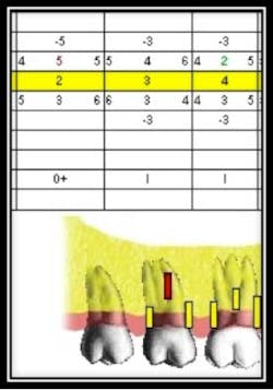
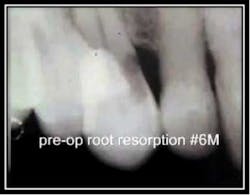
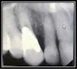
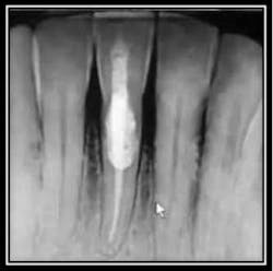
Radiographic Resorption:

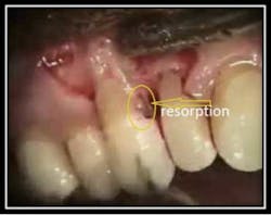
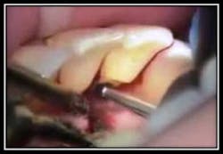
Perioscopic view of external root resorption #15.
Suggested Etiology: Impacted 3rd molar
The below case identifies external root resorption which extends into the cementum and dentinal structures.
#15 reflected in a mirror

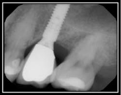
Pre-op radiograph #15

The above pictures identify the resorption. The interior of the resorption is quite hard and may be Reparative dentin, which is formed as a reaction to external stimulation.

Treatment for this type of defect may vary depending upon the extent of the resorption.

This case may be viewed at “External root resorption #15”:
http://www.youtube.com/watch?v=RVzJldSqdXI.

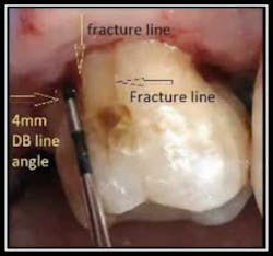
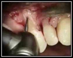
Treatment of Root Resorption by Dr. John Y. Kwan

The below pictures define an area of root resorption that clinically and radiographically exhibited pocketing and bone loss. The attached video link shows a microsurgical repair of root resorption with Geristore; a radiopaque, hybrid ionomer composite used for subgingival restorations.
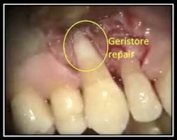
Radiorgraphic resorption MF #6
The extent of resorption is uncovered
Retention preparation to enhance bonding

Geristore repair

Post-op radiograph
3 months Post restorative treatment; healthy tissue The video link “Microsurgical Repair of Root Resorption with Geristore” can be viewed at: http://www.youtube.com/watch?v=x8BD9883xAY.Periodontal Microsurgery Geristore Repair of Root Resorption
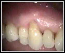
By Dr. John Y. Kwan
The below photos and corresponding video link show a root resorption and Geristore repair of #25 lingual.

A probe defines the resorption

Retention grooves being placed for Geristore placement

Post treatment X-ray
3 months post Geristore repair The video link “Microsurgery Geristore Repair of Root Resorption” may be viewed at:http://www.youtube.com/watch?v=awhZ-_3wfdo.Conclusion: We suggest that the benefits of Perioscopy as a diagnostic tool in the early detection of external root resorptions may aid in the retention of the tooth and help define appropriate therapy based on the extent of the resorption.
Dr. Kwan is a board-certified periodontist and maintains a private practice providing periodontal and implant care in Oakland/Berkeley, Calif. He is an Associate Clinical Professor at the University of California, San Francisco School of Dentistry. Dr. Kwan is also the President/CEO of Perioscopy Incorporated.
Ms. Newkirk graduated from the University of Alaska, Anchorage, in 1981 and has dedicated the last 30 years to excellence in clinical dental hygiene and periodontics. Suzanne is a periodontal therapist, dental hygiene coach and mentor. She is also a Perioscopy instructor and speaker. Ms. Newkirk is a member of the American Dental Hygienists' Association and Washington State Dental Hygienists' Association.

To read articles in RDH eVillage FOCUS written by John Kwan and Suzanne Newkirk, click on Kwan and Newkirk.