Pretreatment England Fo

Subgingival anomalies and pathologies viewed with the Perioscope: root fractures (Part 3)

Jan. 4, 2013
In a third installment, John Kwan, DDS, Robert Gottlieb, DDS, and Suzanne Newkirk, RDH continue the exploration of various anomalies and pathologies visualized with the Perioscope that have been found to be contributing factors in localized periodontal breakdown. Included are three case studies dealing with horizontal and vertical root fractures.

In the abstract “A Review of Root Fractures: Diagnosis, Treatment and Prognosis,” by Malhotra, Kundabala and Acharaya**, root fractures are defined as fractures involving the dentin, cementum and pulp and occur in less than 8 percent of traumatic injuries to permanent teeth.

Root fractures are broadly classified as horizontal (transverse) and vertical root fractures (VRF) and can vary in severity.

Treatment of root fractures depends on a number of factors and is typically determined by the extent of the subgingival fracture, remaining coronal tooth structure, location of the fracture line, pulp vitality and length and morphology of the roots. Frequently, an interdisciplinary or multidisciplinary approach is required for complete rehabilitation of the tooth.

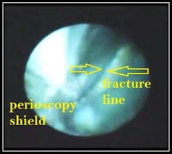
Case #1: Horizontal Root Fracture #9
By Robert Gottlieb, DDS, and Suzanne Newkirk, RDH

This 48-year-old female patient was referred for evaluation of tooth #9, which presented with a 7mm pocket on the straight facial and a 9mm pocket on the mesial.
#9 Pre-treatment photo
#9 Pre-op X-ray Note: Endodontic work #’s 7-10 were done years previously in England. Looking toward the apex of the tooth, the below photo and corresponding video clearly show a horizontal fracture traversing the root.

Root fractures that develop communication between the gingival sulcus and the fracture site have a poor prognosis because of bacterial contamination.

Due to the extent of the above fracture, treatment recommendations for this patient included extraction and immediate implant placement, which the patient proceeded with.

The video “Horizontal Root Fractures: Why Guess about a fracture?” may be viewed at:
http://www.youtube.com/watch?v=t-coebmV6cM

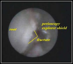
Case #2: Horizontal Hair Line Fracture #19
By Robert Gottlieb, DDS, and Suzanne Newkirk, RDH

Root fractures in posterior teeth usually occur as a result of indirect trauma such as parafunctional habits, traumatic occlusion, extensive tooth decay and iatrogenic causes, and may be symptomatic, or non-symptomatic. This 52-year-old male was referred for a complete periodontal exam. A chief complaint was sensitivity on the lower left to cold, touch and pressure on tooth #19, which had recently had a small occlusal composite placed. The below photo and corresponding video clearly show two hair line vertical fractures connected to a horizontal fracture traversing the buccal surface of tooth #19.
Minor insults (hair line fractures) may lead to concussion injury, or nonvitality. Management of hairline root fractures may include constant observation and vitality tests performed on a regular basis. The video “Hair line root fractures viewed with the Perioscope” may be viewed at:http://www.youtube.com/watch?v=d5vC8vWwhUUCase #3: Vertical Root Fracture #15
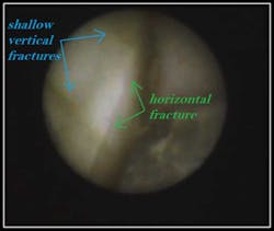
By Dr. John Y. Kwan
Vertical root fractures typically extend through the long axis of the root toward the apex and commonly occur in endodontically treated teeth. Depending upon the severity of the fracture, an interdisciplinary and/or multidisciplinary approach may be required for functional and esthetic rehabilitation of the tooth. Intraosseous fractures typically create deep, narrow, sharply defined isolated periodontal pockets and radiographic examination may reveal unilateral thickening of PDL along the fracture side of the root. If the fracture is not apparent radiographically, diagnostic tests may be considered using a bite-test, transillumination test, or confirmation of the VRF via surgical exploration. Confirmation of root fractures with Perioscopy provides the most minimally invasive, non-surgical method available. The below photo and corresponding video clearly show a vertical root fracture.
The video “Root Fracture #15M: Perioscopy Video” may be viewed at: http://www.youtube.com/watch?v=hk7-R3RJG3w We believe that appropriate management and treatment of root fractures begins with the correct diagnosis. Prior to Perioscopy, confirmation of root fractures was frequently done via exploratory flap surgery. Perioscopy provides the most minimally invasive, non-surgical modality available for confirmation of root fractures. More information on Perioscopy may be found at: http://www.perioscopyinc.com/perioscopy-technology.php ** A Review of Root Fractures: Diagnosis, Treatment and PrognosisDent Update. 2011 Nov;38(9):615-6, 619-20, 623-4 passim. Department of Conservative Dentistry and Endodontics, Manipal College of Dental Sciences, Mangalore, Manipal University, India.http://eprints.manipal.edu/2352/1/A_Review_of_Root_Fractures-_Final_print.pdf
Dr. Kwan is a board-certified periodontist and maintains a private practice providing periodontal and implant care in Oakland/Berkeley, Calif. He is an Associate Clinical Professor at the University of California, San Francisco School of Dentistry. Dr. Kwan is also the President/CEO of Perioscopy Incorporated.
Dr. Bob Gottlieb received his Certificate in Periodontics from the University of Washington. Practicing for 35 years, he was voted one of Seattle’s Best Dentists. Dr. Gottlieb mentors and directs numerous study clubs.
Ms. Newkirk graduated from the University of Alaska, Anchorage, in 1981 and has dedicated the last 30 years to excellence in clinical dental hygiene and periodontics. Suzanne is a periodontal therapist, dental hygiene coach and mentor. She is also a Perioscopy instructor and speaker. Ms. Newkirk is a member of the American Dental Hygienists' Association and Washington State Dental Hygienists' Association. To read articles in RDH eVillage FOCUS written by John Kwan, Robert Gottlieb, and Suzanne Newkirk, click on Kwan, Gottlieb, and Newkirk.