Orthodontic case study: ClearCorrect treatment utilizing extraction of lower left central incisor
For additional information on ClearCorrect, click here.
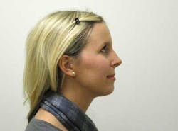
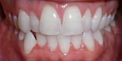
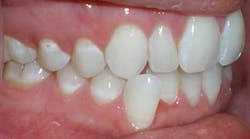
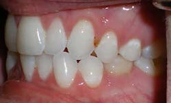
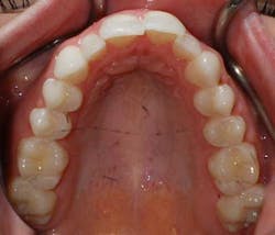
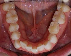
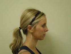
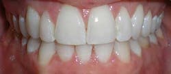
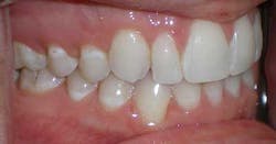
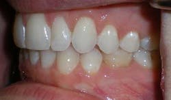
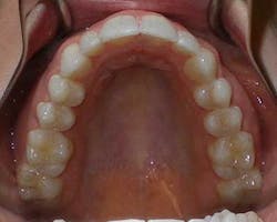
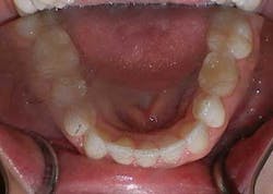
A 27-year-old female presented with a chief complaint of crowding on the upper and lower arches. Upon evaluation, it was noted that the patient had 2 mm and 4.5 mm of maxillary and mandibular arch length deficiency respectively, a missing lower left second bicuspid with primary tooth still present, small upper lateral incisors, cross-bite of the lower right canine, and lower midline shift to the right of 2 mm. (Figs. 1-8)
Figs. 1-8
The patient preferred clear aligner therapy and rejected the treatment plan of comprehensive treatment with full upper and lower fixed appliances. Her general dentist was consulted and the two treatment plan options were agreed upon. It was recommended to the patient that the first option was to align the upper arch and extract the lower left central incisor and use the extraction space to resolve the crowding and cuspid cross-bite on the lower with no IPR on upper and lower. The second option was to open space around the upper laterals to restore to optimal size and IPR and flaring on the lower to resolve crowding. The patient agreed to the first option, but was hesitant about the idea of the extraction.
ADDITIONAL READING |Orthodontic tooth movement with clear aligners: seeing results with the invisible
To ease the patient’s concerns about the extraction space being visible, records were taken. Initial records consisted of intra- and extraoral photos, panoramic radiograph, bite registration, and upper and lower PVS impressions. The records along with the prescription were sent to ClearCorrect with the plan that the incisor would be extracted after the trays were made and received. The patient could then have the extraction and immediately come to the office to receive the trays, which we placed composite in the extraction space pontic that was made into the retainer to mask the space. ClearCorrect was instructed to leave the tooth in the tray and to make it smaller every day as the space closed.
Treatment sequence and detail
The patient was treatment planned for 16 aligners over 10 months of treatment time. The patient was instructed to wear each tray for three weeks at least 22 hours a day. The patient was given a set of trays to wear and a set to take home and change into three weeks later at every appointment. After the trays were completed, the lower right canine did not track well (intruded). To extrude the cuspid, we placed a clear button on the buccal surface of the tooth and used an elastic stretched over the occlusal surface. We connected it to the lingual side of the tray that was adjusted to a point so the elastic had a place to connect to. The patient was instructed to wear the elastic full time (22 hours a day) and remove it only when eating or brushing. A refinement was done with an additional two trays to finish treatment. New upper and lower PVS impressions were taken and sent to ClearCorrect to make the new trays. Engagers were placed on the lower incisors to help control extraction space closure by bodily movement instead of tipping. We used Transbond (3M Unitek, Monrovia, Calif.).
Finishing and retention
As noted above, the patient had a retained primary molar. Since the primary molar had larger mesial-distal dimensions than a second premolar, the completed treatment resulted in an acceptable compromise of interdigitation on this side. Treatment progressed very well, although a refinement was needed to finish the positioning of the lower right cuspid at the end of treatment (Figs. 9-16). The patient was given final upper and lower in-house thermoformed retainers to wear at night. Then a few months later, a permanent lingual retainer was bonded at the patient’s request on the lower from cuspid to cuspid.
Figs. 9-16
Dr. Darren Miller discloses no conflict of interest.


