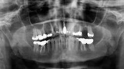
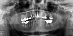
A 71-year-old female is referred for a second opinion by her general dentist. CC: A painful right lower jaw and moderate-to-large growth … Panorex imaging reveals a 2 cm to 3 cm diameter radiolucency that is not well-defined in the right posterior body and ramus of the mandible …
This article first appeared in the newsletter, DE's Breakthrough Clinical with Stacey Simmons, DDS. Subscribe here.
A 71-year-old white female is referred to the office for a second opinion by her general dentist. CC: A painful right lower jaw and moderate-to-large growth in the right posterior mandible behind tooth No. 31. The patient states that the “lump” in her right posterior mandible began to grow only two weeks ago, although the pain in her right jaw has been present for about six weeks, and began with some numbness in her right lip. The consistence of the pain in her right jaw is constant and throbbing. She has already been to one surgical specialist prior to the mass forming. The specialist performed a clinical exam and x-ray imaging, but could not find any abnormalities. He referred the patient on to testing for trigeminal neuralgia.
The patient’s medical history is significant for morbid obesity, high cholesterol, hypertension, coronary artery disease, and endometrial carcinoma 14 months ago that was treated with radiation and chemotherapy. She had an appointment two months ago with the oncologist and stated that everything was going well.
While performing a clinical exam, a 2.5 cm diameter round, greyish-pink mass is observed directly behind tooth No. 31. There is no tenderness to palpation, although the tissue is friable and bleeds easily. Tooth No. 31 is mobile (Class II), with little tenderness to palpation. The other teeth in that quadrant are normal. The rest of the intraoral exam is normal. There is no extraoral swelling, no bony expansion to palpation of the mandible, and no cervical lymphadenopathy. The right mandibular division of the trigeminal nerve shows paresthesia upon nerve testing.
Panorex imaging reveals a 2 cm to 3 cm diameter radiolucency that is not well-defined in the right posterior body and ramus of the mandible around the region just anterior to the mandibular foramen. The lucency seems to extend to tooth No. 31.
What is the next appropriate step to take with this patient? What is your differential diagnosis?
Send your answers to [email protected] or join our Facebook group to discuss this oral pathology case and more. Next month we will discuss the final diagnosis and recommended treatment for this case.
This article first appeared in the newsletter, DE's Breakthrough Clinical with Stacey Simmons, DDS. Subscribe here.
For more pathology articles, click here.
Do you have an interesting oral pathology case you would like to share with Breakthrough’s readers? If so, submit a clinical radiograph or high-resolution photograph, patient history, diagnosis, and treatment rendered to: [email protected]. We will let you know if we select your case!


