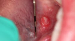
Dr. Stacey Simmons gives her definitive diagnosis and treatment for the oral pathology case of an 8-year-old female who presents with a raised, round soft-tissue lesion under the left side of her tongue, along the sublingual salivary glands.
This article first appeared in the newsletter, DE's Breakthrough Clinical with Stacey Simmons, DDS. Subscribe here.
Last month I described the oral pathology case of a healthy 8-year-old female who presented for her routine recare visit. While performing a head, neck, and oral cancer screening exam, I observed a raised, round soft-tissue lesion under the left side of the tongue along the sublingual salivary glands. The lesion was approximately 6 mm x 6 mm; its center was white with a red outside border. Neither the lesion nor area in general were tender to palpation, and both mom and patient were unaware of its presence. There was no recollection of trauma to the area.
Differentials
- Aphthous ulcer (minor) aka canker sore
- Blockage of salivary duct (mucocele)
- Idiopathic irritation/traumatic ulcer
- Recurrent intraoral herpes simplex (RIHS)
Definitive diagnosis: Aphthous ulcer (minor)
Apthus ulcers (minor) are “small superficial ulcers of the oral gland-bearing mucosa that occur episodically in clusters of one to five lesions.” (1) It is the most common type of aphthous ulcer found. (2) The lesions occur throughout the mouth, including the soft palate, floor of mouth, tongue, mucobuccal fold, buccal mucosa, lips, and movable mucosa (nonkeratinized). (2)
Etiology and treatment
Aphthous ulcers have a slight prevalence in women (2) and treatment is palliative, as the lesions are self-limiting and will resolve in typically seven to 10 days. Application of rinses or topical ointments can be applied; numbing agents such as benzocaine are also helpful.
Of interest, the recurrence (and appearance) of aphthous ulcers is similar to RIHS. One of the main differentiating features is that RIHS is typically found on keratinized tissue, and the lesions often return to the same location. It is, therefore, prudent to be aware of patterns and frequency of any lesions that have a common occurrence.
In this particular case, the patient was seen 10 days later, and the lesion had self-resolved. A discussion with mom revealed that the patient did indeed have a tendency to get aphthous ulcers. This lesion was apparently small and isolated enough (as a single lesion) that the discomfort factor was not an issue. No further follow-up was necessary.
References
1. Sapp JP, Eversole LR, Wysocki GP. Contemporary Oral and Maxillofacial Pathology. St. Louis: MO: Mosby; 1997:246-247.
2. Wood NK, Goaz PW. Differential Diagnosis of Oral and Maxillofacial Lesions. 5th ed. Maryland Heights, MO: Mosby Publishing; 1997:165-167.
This article first appeared in the newsletter, DE's Breakthrough Clinical with Stacey Simmons, DDS. Subscribe here.


