For years, Melinda Harr, DDS, was frustrated by areas that appeared "suspicious" around amalgams and under sealants. Then, she discovered a caries detection device that would help her to identify caries in these situations and more easily determine whether a restoration would be required.
For years, I was frustrated by areas that appeared “suspicious” under sealants and around older amalgams. Radiographs were usually of little help diagnosing caries in these situations. During crown preps, I often discovered that these gray suspicious areas at the margins of old amalgam restorations were decay. I was concerned about how many of these areas I had been failing to diagnose.
Caries detection devices have always been of interest to me. Our office had tried one, but it had limitations. Then, in early 2015 at the Minnesota Star of the North dental conference, I heard great things about the Canary System, a laser-based caries detection system from Quantum Dental Technologies, during one of the seminars. I was impressed that it could detect caries under sealants, between teeth, and around existing restoration margins. I purchased the Canary System at the conference and have been using it ever since.
The Canary gives me more confidence when diagnosing. If an area looks questionable, we use the Canary System in hygiene to verify the diagnosis. In some instances, I ask my dental assistant to check any suspicious areas at the restorative appointment while the anesthetic is taking effect. If we determine that additional treatment is needed, we offer to complete it the same day.
We derive our treatment decisions from various factors, including tooth surface, Canary Number, patient risk factors, and clinical judgment. The Canary Number is the score a tooth gets on the Canary Scale after being scanned (figure 1). Canary Numbers above 20 indicate decay. Previously, I would question if suspicious areas I observed in interproximal areas on digital radiographs were advanced enough to require restorations. On an interproximal surface, a Canary Number above 20 generally warrants a restoration.
Figure 1: The Canary Scale. Canary Numbers from 0–20 indicate healthy tooth structure, whereas a score of 21–70 indicates decay and a score of 71–100 indicates advanced decay.
In this case, the Canary reading on tooth No. 3 was 32 from the distal-interproximal (figure 2). The caries had just begun to extend into the dentin, yet we discovered it early enough to keep the restoration conservative and preserve a significant amount of healthy enamel. We have recommended restoring other interproximal areas.
Figure 2: The Canary reading on tooth No. 3 was 32 from the distal-interproximal. The caries had just begun to extend into the dentin.
In another case, the Canary System confirmed that an existing composite on tooth No. 18 had mesial and occlusal caries with Canary Numbers 38 and 43, respectively (figure 3).
Figure 3: Tooth No. 18 had mesial and occlusal caries with Canary Numbers 38 and 43, respectively.
The system has made me more confident and comfortable saying a restoration should be placed or that we should monitor the area. The information obtained from the Canary System has been accurate, and we have not had any false positives.
We share the photos taken during caries excavation with our patients to document the caries. The patient response has been overwhelmingly positive. They appreciate the technology that we have available to improve the accuracy of our exams and treatment planning.
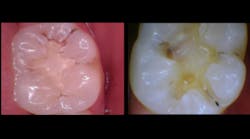
Occasionally, new patients become concerned if we discover restorations are needed after checking the teeth with the Canary System but their previous dentists had not found any caries. During one new patient visit, the clinical exam demonstrated a typical virgin pit and fissure (figure 4). The Canary Number was 52, and excavation revealed caries well into the dentin that was not evident on the radiograph.
Figure 4: The clinical exam demonstrated a typical virgin pit and fissure; however, the tooth's Canary Number was 52, and excavation revealed caries well into the dentin that was not evident on the radiograph.
The Canary System improves patients' understanding of their oral health and helps eliminate patient concerns by serving as an education and communication tool that is extremely valuable during the exam process.
In summary, the pros of the Canary System outweigh its cons by far. It has helped me clearly identify the areas that need to be restored and has reduced uncertainty when diagnosing. Patients love the fact that we are able to catch the cavities earlier to prevent discomfort and minimize loss of tooth structure. And the minimal time it takes during the exam appointments greatly increases the quality of my dentistry and improves my peace of mind.
Editor's note: This article first appeared in Pearls for Your Practice: The Product Navigator. Click here to subscribe. Click here to submit a products article for consideration.


