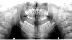
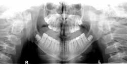
Dr. R.F. John Holtzen gives his diagnosis for the oral pathology case of a 14-year-old male who was referred to evaluate his lower third molars, which appeared on panoramic radiograph to have significant well-circumscribed, expansile radiolucencies around the impacted teeth. The medical history was remarkable for mental delay and recent excision of a basal cell carcinoma from the patient's chest.
Editor's note: This article first appeared in DE's Breakthrough Clinical with Stacey Simmons, DDS. Find out more about it and subscribe here.
Last month we described the case of a 14-year-old male who had been referred to the oral and maxillofacial surgeon to evaluate his lower third molars, each of which appeared on panoramic radiograph to have significant well-circumscribed, expansile radiolucencies around the impacted teeth. The patient's medical history was remarkable for mental delay and recent excision of a basal cell carcinoma from the patient's chest.
Removal of the lower third molars and the associated cystic lesions produced an expected histopathologic diagnosis of bilateral keratocystic odontogenic tumors. Because of the high suspicion of this diagnosis, the surgery included aggressive removal of the lesions with peripheral ostectomy and treatment with Carnoy's solution.
The patient was determined to have Gorlin syndrome, which affects approximately 1 in 31,000 people. Often referred to as nevoid basal cell carcinoma syndrome, individuals suffering with this syndrome often have significant mental disability; macrocephaly; skeletal abnormalities of the spine, ribs, and skull; and pits in the palms of the hands and soles of the feet.
Affected individuals will be a high risk for development of multiple keratocystic odontogenic tumors starting in adolescence and continuing into their thirties, and will require continuous observation and management for this issue. They are also at higher risk for various types of cancer, most significantly basal cell carcinomas of the face, chest, and back.
Editor's note: This article first appeared in DE's Breakthrough Clinical with Stacey Simmons, DDS. Find out more about it and subscribe here.


