A healthy 40-year-old male presents with a fluctuant mass approximately 12 mm x 24 mm on the lingual side of the left mandibular jaw, along the palatoglossal arch muscle. The mass is tender to palpation. Both the diagnosis and genesis for the origin of this soft-tissue mass are surprising. After reading the details of this oral pathology case, how would you proceed with treatment?
Editor's note: This article first appeared in DE's Breakthrough Clinical with Stacey Simmons, DDS. Find out more about the clinical specialties newsletter created just for dentists, and subscribe here.
Presentation and chief complaint
A healthy 40-year-old male presents to the office with this chief complaint:
“I have some swelling on the lower left side toward the back of my mouth on my jawbone. It’s been there for two days and seems to be getting worse."
Clinical exam
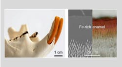
Clinical assessment reveals a fluctuant mass that is tender to palpation on the lingual side of the left mandibular jaw, inferior and lingual to where No. 17 would be, along the palatoglossal arch muscle. The size of the mass is approximately 12 mm x 24 mm, the tissue in the surrounding area is normal in color, and there is no opposing dentition or history of trauma to the area (figures 1 and 2). Upon initial assessment, the panoramic radiograph is insignificant (figure 3).


Figure 3
With this information, how would you proceed with treatment for this patient? I will tell you that both the diagnosis and genesis for the origin of this soft-tissue mass are surprising. Further, multiple complications have occurred while rendering treatment.
Send your answers to [email protected] or join our Facebook group to discuss this oral pathology case and more. Next month, we will discuss the final diagnosis and recommended treatment for this case.
Do you have an interesting oral pathology case you would like to share with Breakthrough’s readers? If so, submit a clinical radiograph or high-resolution photograph, a patient history, diagnosis, and treatment rendered to: [email protected]. We will let you know if we select your case.
Editor's note: This article first appeared in DE's Breakthrough Clinical with Stacey Simmons, DDS. Find out more about the clinical specialties newsletter created just for dentists, and subscribe here.