What is caries? This might seem a simple question, but the answer will help direct us to improving how we detect, monitor, and treat this common oral disease. In this article, we will examine a definition of caries, the recent FDI World Dental Federation statement on caries, and the benefits of selected modern methods of caries management and prevention.
Caries defined
In 2001, the National Institutes of Health Consensus Conference on the Diagnosis and Management of Dental Caries Throughout Life concluded the following: “Dental caries is an infectious, communicable disease resulting in destruction of tooth structure by acid-forming bacteria found in dental plaque, an intraoral biofilm, in the presence of sugar. The infection results in the loss of tooth minerals that begins with the outer surface of the tooth and can progress through the dentin to the pulp, ultimately compromising the vitality of the tooth.”1
If caries is a disease that results in the destruction of tooth structure, then we need to find
- the right tools to detect and monitor changes in caries lesions;
- the right tools or products to treat the early stages of this disease process; and
- a means of engaging patients in this process so that they can monitor their progress.
It is important to emphasize that restorations do not treat the disease. They only treat the effects of the disease once a lesion has grown to involve the dentin.
Latest FDI World Dental Federation goals
In August 2017, the Caries Prevention and Management Chairside Guide published by FDI World Dental Federation (figure 1) summarized goals related to caries as follows: "The goal is to reduce the impact of caries development by intervening as soon as possible to manage further tooth destruction, and reversing the caries process in favour of remineralization. Ideally, the management of early caries lesions should involve the least invasive approach capable of preventing disease progression and empowering the patient to improve and maintain their own oral health."2

Figure 1:Caries Prevention and Management Chairside Guide
The first step is to detect and monitor caries. The FDI's Chairside Guide further states: "The essential challenge is to differentiate between firstly a lesion which is active today and continuing to suffer net loss of mineral, with demineralization being out of balance with remineralization, as opposed to a lesion of similar severity which has been switched off and becomes inactive, i.e., arrested or remineralized."2
In essence, we need to find a system that can detect and monitor the changes in the crystal structure of the tooth (enamel, dentin, and cementum). Bacterial activity may have some correlation, but it is the changes in the structure of the lesion that are the end result of the caries process.
Traditional methods
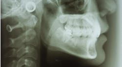
Traditionally, we have relied upon visual examination and radiographs for this process. However, the literature shows that radiographs are limited:
- Dove found that "overall the strength of the evidence for radiographic methods for the detection of dental caries is poor for all types of lesions on proximal and occlusal surfaces."3 The review went on to conclude that radiographs are "beneficial only if the intervention is the surgical removal of tooth structure and detrimental if it is used for non-invasive remineralization methods."3
- In their review of radiographic diagnostic procedures, Pretty and Maupome concluded that "for interproximal lesions a clinician using radiographs can be very certain of the lack of disease in apparently sound surfaces (97% specificity) but not as certain that disease is indeed present in suspect interproximal surfaces (54% sensitivity)."4
- A study in 2010 using bitewing radiographs for detection of interproximal caries found 10.6% of enamel caries, 17.8% of dentine caries, and 40.2% of deep dentine caries. This indicated that, at best, bitewing radiographs could detect deep lesions less than 50% of the time.5 This low sensitivity for detection of enamel lesions in interproximal regions is not unusual and may be due to the irregular shape and low contrast of these small early lesions.6
Radiographs and visual examination are valid diagnostic tools for the detection of larger lesions,7,8 but there is need for more sensitive methods that can detect, measure, and monitor lesions. X-rays and visual exams can't provide these functions.
International Caries Detection and Assessment System II
The International Caries Detection and Assessment System II (ICDAS), which is a visual ranking system listed in the FDI's Chairside Guide, can provide a more robust system for ranking (figure 2).1 The system uses six clinical conditions for monitoring caries. Early lesions are measured by looking at surface stain on wet or dry surfaces.

Figure 2: ICDAS II lesion ranking
Research has shown that ICDAS presents good reproducibility and accuracy for in vitro and in vivo detection of primary caries lesions at different stages of the disease,11,12,13 but it can’t measure subtle changes in caries lesions in response to various preventive or remineralization therapies.
Modern methods of caries detection
Three different approaches to caries detection on the market today are:
- fluorescence
- transillumination
- frequency domain photothermal radiometry and modulated luminescence (PTR-LUM)
Fluorescence
Fluorescence is simply the emission of light from an object that has absorbed light at a specific wavelength. This is the core technology in Soprolife (Acteon), Spectra (Air Techniques), and Diagnodent (KaVo Kerr Group).14 These devices produce a glow from the tooth surface when an LED or laser light is shined on the tooth. The literature indicates that the glow or fluorescence is from one or more of the following 14–20
- bacterial porphyrins (bacterial breakdown products)17
- stain
- tartar
- food debris
Soprolife and Spectra claim that caries exhibits red fluorescence when excited by the wavelengths of light emitted by these devices.
Transillumination
Transillumination involves shining either visible light or near-infrared light through a tooth and measuring the scatter or disruption of the light. Sound enamel is composed of densely packed hydroxyapatite crystals that allow light to pass through them. When demineralization occurs, the light is disrupted and the area will appear as a shadow. Shadows may indicate caries is present because demineralized areas of enamel or dentine scatter light more than sound areas. Therefore, caries appear as darker areas.
Frequency domain photothermal radiometry and modulated luminescence (PTR-LUM)
Photothermal radiometry and modulated luminescence (PTR-LUM) uses energy conversion technology to examine and create an image of a tooth. Pulses of laser light are aimed at a tooth, and the light is then converted to heat (photothermal radiometry or PTR) and light (luminescence or LUM), which are emitted from the tooth surface in response to the modulated pulses. These pulses of laser light enable the clinician to examine lesions up to 5 mm below the surface.8,9,10
Caries modifies the thermal properties and luminescence of healthy teeth. As a lesion grows, there is a corresponding change in the PTR-LUM response signal. In effect, the heat confined to the region with crystalline disintegration (caries) increases the PTR and decreases the LUM response signal. As remineralization progresses and enamel prisms start to reform their structure, the thermal and luminescence properties begin to revert towards those of healthy tooth structure.11-14 The system detects very small changes in heat (less than 1–2 degrees Celsius), much less than that generated by a dental curing light.
The Canary System (Quantum Dental Technologies) uses a low-power laser diode (<45 mW at the tooth surface) at 660 nm and modulated at 2 Hz.15 Research has demonstrated that Canary’s energy conversion technology can be harnessed to help oral health professionals detect and diagnose:- Lesions and defects £ 5 mm below the enamel surface16-19
- Occlusal pit and fissure caries9,19-21
- Smooth surface caries11,22,23
- Acid-erosion lesions8,24-27
- Root caries28,29
- Interproximal caries lesions10,30-34
- Caries beneath fissure sealants35-37
- Caries around margins of restorations and crowns38-40
- Caries beneath the intact margins of composite resins41
- Caries beneath the intact margins of amalgam restorations42
- Demineralization and remineralization of early lesions14,29,43-45
- Caries beneath clear resin infiltrants46
- Caries around orthodontic brackets47
The earliest visual clinical sign of dental caries is the "white spot lesion." When this is first seen, the carious process has been going on for months. Figure 3 shows a cross-section of a white spot lesion.

Figure 3: Structure of white spot lesion
Even though the surface appears intact, the lesion is at least 540 microns in depth. In this case, scanning with The Canary System indicates that a lesion is present. These early lesions can be treated before cavitation, and they are amenable to remineralization.48,49 The key is to find the lesion and use technology to monitor the changes in the lesion as it undergoes remineralization.
Patient engagement
Patient engagement is critical for the success of any oral health program.50-52 In treating caries, patients understand the need to brush and floss teeth, but they have no way to gauge if this is making a difference. Caries imaging, especially using radiographs for imaging proximal lesions, is difficult for patients to understand as they are looking a small black shadow on an image.
Spectra and Sopro capture images of the surface under examination. The fluorescence from the tooth surface is translated into colors which indicate the degree of porphyrin entrapment on the tooth surface. The Canary System has a voice that provides the Canary Number after each four-second scan. This helps both the operator and the patient to understand what is being measured. The Canary System contains an intraoral camera so images of the surface being examined can be shown to the patient. Using the detail scan mode, the Canary Numbers are recorded on the image and a report can be generated for the patient showing the Canary Numbers and treatment recommendations.
Case study: Measuring lesions over time
The major challenge in clinical practice is how or can we track changes in lesion depth or volume over time. In Figure 4 we see two brown spots on the buccal surface of a mandibular molar.

Figure 4: Is this lesion growing or recrystalizing?
We started this patient on a preventive/remineralization program in our clinical practice in December 2012 that involved the application of fluoride varnish (Vanish Fluoride Varnish, 3M ESPE) every three months and brushing with Clinpro 5000 Toothpaste (3M ESPE) twice daily. We monitored the lesions with The Canary System. By January 2014, the lesions still visually appeared unchanged. We probed the surfaces with the back of an explorer. They were harder, and The Canary System measurements at the center of each lesion showed a drop in Canary Number into the healthy zone. Visual examination does provide some information, but one needs devices that can measure changes below the tooth surface.
Summary
Caries is a disease that results in the destruction of the crystal structure of the tooth. Treating caries does not involve the placement of restorations, but the detection, management, and monitoring of the lesion over time. There are a number of caries-detection devices on market, but the critical questions are:
- What are they detecting?
- Can they monitor changes in the lesion volume?
- How do they engage patients in understanding the disease process?
- Can these devices detect demineralization and remineralization of the lesion on all tooth surfaces, including pits and fissures and beneath restoration margins?
Oral health care providers are now able to detect caries early in the disease process using the appropriate caries detection device. We now need to incorporate the proper device into our clinical practice and start to treat caries as soon as the lesions appear. Treatment may not be placement of a restoration, but may involve a sealant, a remineralization therapy, or even products that will act as a scaffold to initiate enamel remineralization. Modern treatment of caries is moving away from placement of restorations to the detection and management of lesions over time with a wide range of therapies.
Disclosure: Stephen Abrams, DDS, is the CEO and cofounder of Quantum Dental Technologies, which has developed The Canary System mentioned in this article. He has not received any compensation for the preparation of this article.
References
1. Mjör, I. Clinical diagnosis of recurrent caries. Journal of the American Dental Association. 2005;136(10):1426-1433.
2. Photonics for Health Care. In Handbook of Biophotonics Volume 2. Popp J, Chiou A, Heinemann SH eds. 2012. Wiley-VCH: Weinheim, Germany.
3. Lennon AM, Brune L, Zimmermann O, Gross U, Attin T. The ability of selected oral microorganisms to emit red fluorescence. Caries Res. 2006;40(1):2-5.
4. Coulthwaite L, Smith PW, Higham SM, Verran J. The microbiological origin of fluorescence observed in plaque on dentures during QLF analysis.Caries Res. 2006;40(2):112-116.
5. Thomas R, van der Mei H, van der Veen M, de Soet JJ, Huysmans M. Bacterial composition and red fluorescence of plaque in relation to primary and secondary caries next to composite: an in situ study. Oral Microbiol Immunol. 2008;23(1): 7-13.
6. König K, Sigusch B, Glockmann E, Eick S, Pfister W. Red light kills bacteria via photodynamic action. Cell Mol Biol. 2000;46(7):1297-1303.
7. Pretty IA. Caries detection and diagnosis: novel technologies. J Dent. 2006;34(10):727-739.
8. Jeon RJ, Phan T, Wu A, Kulkarni G, Abrams S, Mandelis A. Photothermal radiometric quantitative detection of the different degrees of demineralization of dental enamel by acid etching.J Physique IV France. 2005;125: 721–772.
9. Jeon, R.J., et al., Diagnosis of pit and fissure caries using frequency-domain infrared photothermal radiometry and modulated laser luminescence. Caries Res, 2004. 38(6): p. 497-513.
10. Jeon, R.J., et al., Detection of interproximal demineralized lesions on human teeth in vitro using frequency-domain infrared photothermal radiometry and modulated luminescence. J Biomed Opt, 2007. 12(3): p. 034028.
11. Matvienko, A., Jeon, R. J., Mandelis, A., Abrams, S. H., Amaechi, B. T, Photothermal detection of incipient dental caries: experiment and modeling. XVI International Conference on Photoacoustic and Photothermal Phenomena (ICPPP16), 2011.
12. Jeon, J.G., et al., Experimental Investigation of Demineralization and Remineralization of Human Teeth Using Infrared Photothermal Radiometry and Modulated Luminescence. Proc SPIE, 2008. 6856: p. 68560B.
13. Matvienko, A., A. Mandelis, and S. Abrams, Robust multiparameter method of evaluating the optical and thermal properties of a layered tissue structure using photothermal radiometry. Appl Opt, 2009. 48(17): p. 3192-203.
14. Silvertown, J.D., et al., Remineralization of natural early caries lesions in vitro by P11 -4 monitored with photothermal radiometry and luminescence. J Investig Clin Dent, 2017.
15. Jeon, R.J., et al., Dental diagnostic clinical instrument ("Canary") development using photothermal radiometry and modulated luminescence. Journal of Physics: Conference Series, 2010. 214: p. 012023.
16. Jeon, R.J., et al., Nonintrusive, noncontacting frequency-domain photothermal radiometry and luminescence depth profilometry of carious and artificial subsurface lesions in human teeth. J Biomed Opt, 2004. 9(4): p. 804-19.
17. Wong, B., Abrams, S.H., Sivagurunathan, K., Silvertown, J.D., Hellen, A., Mandelis, A., Hellen, W.M.P., Elman, G.I., Amaechi, B.T, Correlation with caries lesion depth of The Canary System, DIAGNOdent and ICDAS II, in 60th Annual European Organization for Caries Research Conference 2013, Caries Research Liverpool, UK. p. 433-531.
18. Carey, C., Coleman, S.S. PLM validation of WSL assessment by photothermal radiometry- modulated luminescence technology. in 2014 AADR/CADR Annual Meeting 2014. Charlotte, North Carolina Journal of Dental Research
19. Abrams, S.H., Sivagurunathan, K., Silvertown, J. D., Wong, B., Hellen, A., Mandelis, A., Hellen, W. M. P., Elman, G. I., Mathew, S. K., Mensinkai, P. K., Amaechi, B. T., Correlation with Caries Lesion Depth of The Canary System, DIAGNOdent and ICDAS II. The Open Dentistry Journal, 2017. 11: p. 679-689.
20. Jeon, R.J., et al., Dental depth profilometric diagnosis of pit & fissure caries using frequency-domain infrared photothermal radiometry and modulated laser luminescence. Journal de Physique IV (Proceedings), 2005. 125: p. 741-744.
21. Jeon, R.J., Han, C., Mandelis, A., Sanchez, V., Abrams, S., Dental depth profilometric diagnosis of pit and fissure caries using frequency-domain infrared photothermal radiometry and modulated laser luminescence, in Proceedings of the 6th Annual Indiana Conference G.K. Stookey, Editor. 2003: Indiana School of Dentistry Indianapolis Indiana. p. 49-67.
22. Jeon, R.J., Mandelis, A., Abrams, S., Depth profilometric case studies in caries diagnostics of human teeth using modulated laser radiometry and luminescence. Review of Scientific Instruments, 2003. 74(1): p. 380.
23. Wong, B., Sivagurunathan, K., Silvertown, J.D., Hellen, W.M., Elman, G., Okoye, L.O., Abrams, S.H., Amaechi, B.T, A comparison of methods for the detection of smooth caries, in IADR/AADR/CADR General Session & Exhibition 2015, Journal of Dental Research Boston Massachusetts p. 0305.
24. Sivagurunathan, K., Hellen, A., Silvertown, J.D., Wong, B., Jeon, R.J., Amaechi, B.T., Abrams, S.H., Detection, monitoring and imaging dental erosion with The Canary Lab, in International Association of Dental Research (IADR) 91st General Session. 2013, J Dent Res: Seattle, WA. p. 2901.
25. Abrams, S.H., Matvienko, A., Ye, V., Mandelis, A., Ramalingam K., Amaechi, B. T., Detection and monitoring of dental erosion using PTR-LUM, in IADR/AADR/CADR 89th General Session. 2011, J. Dent. Res.: San Diego, CA p. 238.
26. Pier, S., Lee, H., Carey, C.M. Detection of surface erosion: a novel application for PTR-LUM technology. in Rocky Mountain Dental Conference. 2015.
27. Matvienko, A., Mandelis, A., Abrams, S. H., Amaechi, B. T. Study of Dental Erosion using the PTR-LUM Technique. in XVI International Conference on Photoacoustic and Photothermal Phenomena (ICPPP16). 2011.
28. Jeon, R.J., et al., In vitro detection and quantification of enamel and root caries using infrared photothermal radiometry and modulated luminescence. J Biomed Opt, 2008. 13(3): p. 034025.
29. Jeon, R.J., Hellen, A., Matvienko, A., Mandelis, A., Abrams, S. H., Amaechi, B. T, Detection of demineralized-remineralized lesions on root and enamel of human teeth in vitro using infrared photothermal radiometry and modulated luminescence. Caries Research, 2007. 41: p. 323.
30. Jeon, R.J., et al., Interproximal dental caries detection using Photothermal Radiometry (PTR) and Modulated Luminescence (LUM). The European Physical Journal Special Topics, 2008. 153(1): p. 467-469.
31. Mandelis, A., et al., Dental biothermophotonics: How photothermal methods are winning the race with X-rays for dental caries diagnostic needs of clinical dentistry. The European Physical Journal Special Topics, 2008. 153(1): p. 449-454.
32. Wong, B., Abrams, S.H., Tasevski, C., Sivagurunathan, K., Silvertown, J.D., Hellen, W.H., Elman, G., Amaechi, B.T, Detection of interproximal caries in vitro using The Canary System. J Dent Res, 2014. 93(Spec Iss A).
33. Jan, J., et al., Proximal caries lesion detection using the Canary Caries Detection System: an in vitro study. J Investig Clin Dent, 2016. 7(4): p. 383-390.
34. Uzamere, E.O., Jan, J., Bakar, W.W., Mathews, S.M., Amaechi, B, Clinical trial of the Canary System for proximal caries detection. J Dent Res, 2015. 94 (Spec Iss A).
35. Wong, B., Abrams, S.H., Sivagurunathan, K., Jeon, R.J., Silvertown, J.D., Hellen, A., Mandelis, A., Hellen, W.M.P., Elman, G.I., Ramalingam, K., Ccahuana-Vasquez, R.A., Amaechi, B.T., In vitro detection of caries beneath dental sealant with The Canary System, in 59th ORCA Congress. 2012, Caries Res: Cabo Frio, Brazil p. 268-338.
36.Abrams, S.H., Wong, B., Sivagurunathan, K.S., Jeon, R.J., Silvertown, J.D., Hellen, A., Mandelis, A., Hellen, W.M., Elman, G.I., Ramalingam, K., Ccahuana-Vasquez, R.A., Amaechi, B.T Effect of placing an opaque sealant on Canary Number readings, in International Association of Dental Research 90th General Session. 2012, J Dent Res: Iguaçu Falls, Brazil. p. 7.
37. Silvertown, J.D., et al., Comparison of The Canary System and DIAGNOdent for the in vitro detection of caries under opaque dental sealants. J Investig Clin Dent, 2016.
38. Kim, J.M., A., Matvienko, A., Abrams, S., Amaechi, B. T., Detection of Dental Secondary Caries Using Frequency-Domain Infrared Photothermal Radiometry (PTR) and Modulated Luminescence (LUM). International Journal of Thermophysics, 2012. 33(10-11): p. 1778-1786.
39. Wong, B., Abrams, S.H., Silvertown, J.D., Sivagurunathan, K., Klausz, R., Mandelis, A., Amaechi, B.T, Detection of caries around ceramic crown restorations with The Canary System and DIAGNOdent, in 60th Annual ORCA Congress. 2013, Caries Res Liverpool UK. p. 433-531.
40. Carey, C.M., Coleman, S.S, Antatomy of secondary caries: the early stages. Dent Mat, 2013. 29(Suppl 1): p. e36.
41. Abrams, S.H., Silvertown, J.D., Wong, B., Sivagurunathan, K.S., Jeon, R.J., Mandelis, A., Hellen, W.M.P., Elman, G.I., Amaechi, B.T, Detection of caries around restorations with The Canary System, in International Association of Dental Research 90th General Session. 2012, J Dent Res: Iguaçu Falls, Brazil. p. 1824.
42. Abrams, T.E., Abrams, S. H., Sivagurunathan, K., Silvertown, J. D., Hellen,W. M. P., Elman, G. I., Amaechi, B. T., In Vitro Detection of Caries Around Amalgam Restorations Using Four Different Modalities. The Open Dentistry Journal, 2017. 11: p. 609-620.
43. Hellen, A., et al., Quantitative evaluation of the kinetics of human enamel simulated caries using photothermal radiometry and modulated luminescence. J Biomed Opt, 2011. 16(7): p. 071406.
44. Hellen, A., et al., Quantitative remineralization evolution kinetics of artificially demineralized human enamel using photothermal radiometry and modulated luminescence. J Biophotonics, 2011. 4(11-12): p. 788-804.
45. Wong, B., Silvertown, J.D., Abrams, S.H., Sivagurunathan, K., Amaechi, B.T Detection of remineralization of early caries with The Canary System. in 2014 AADR/CADR Annual Meeting 2014. Charlotte, North Carolina J Dent Res.
46. Wong B, Abrams S, Silvertown J, Sivagurunathan K, Amaechi BT, Hohnk HD. Using the Canary System to evaluate the resistance of resin infiltration to demineralization. In European Organization for Caries Research 62nd Annual Conference. 2015, Caries Research Brussels Belgium: 297-369.
47. Dorfman JM. Cement composition effects on enamel demineralization adjacent to orthodontic brackets: An in vitro study using the canary system. Oral Biology. 2017 Temple University ProQuest Dissertations & Theses Global. (1951782587). p. 54.
48. Gorton J. In vivo inhibition of demineralization around orthodontic brackets. Am J Orthod Dentofacial Orthop. 2003;123(1):10-14.
49. Iijima Y. Early detection of white spot lesions with digital camera and remineralization therapy. Aust Dent J. 2008;53(3):274-280.
50. Edelstein BL, Ng MW. Chronic Disease Management Strategies of Early Childhood Caries: Support from the Medical and Dental Literature. Pediatr Dent. 2015;37(3):281-287.
51. Edelstein BL. Pediatric Dental-Focused Interprofessional Interventions: Rethinking Early Childhood Oral Health Management. Dent Clin North Am. 2017;61(3):589-606.
52. Ng MW, Fida Z. Dental Hygienist-Led Chronic Disease Management System to Control Early Childhood Caries. J Evid Based Dent Pract. 2016;16:20-33.