DIAGNOdent

Caries, a communicable, infectious disease transmitted from adults to children, is one of the most prevalent diseases among people in the United States. Among young people age 17 and younger, 80 percent develop decay. As young as two- to four-years-old, a startling 20 percent of children already have tooth decay.1 In a large survey of the U.S. population more than 15 years ago, the average number of decayed, missing, or filled tooth surfaces (DMFS) averaged 43 for adults aged 35 to 44, rising to 88.7 for adults aged 65 to 74.2
Until recently, early detection of decay by dental professionals has been limited. Many of the diagnostic cornerstones for caries detection taught in dental school — mirror/observation, explorer, and radiographs — are modestly adequate and highly subjective, with low sensitivity. Using currently accepted decay-detection methods results in correct identification on average 17 to 42 percent of the time. For example, use of an explorer alone for caries detection has never been tested scientifically, much less validated.3 However, a more current diagnostic method — the DIAGNOdent (KaVo America) — has been scientifically proven to be more specific and reliable than those previously mentioned detection methods.4
The process of tooth decay requires bacteria, most commonly Streptococci mutans and Lactobacillus acidophilus, and a fermentable carbohydrate. In combination they produce acid by-products. These acid by-products penetrate the tooth enamel or root surfaces, rapidly dissolving through channels into the calcium phosphate enamel surface known as carbonated hydroxyapatite.5 Demineralization is the surface interruption of the penetrating acids. Clinically, cavitation is the critical point when the penetrated enamel layer would require restorative intervention. The current American diet is changing, resulting in increased sugar consumption, more fast food meal choices, and a variety of ingested acids that are transported via sports drinks and carbonated beverages that intensify the decay process.
Remineralization is the chemical process in reverse. The carbonated hydroxyapatite crystals utilize the protective factors in saliva and the oral cavity. These include minerals such as fluoride, calcium, phosphate, and antibacterials that can resist acid challenges. These can re-form enamel with a more dense, resilient tooth mineral that is less soluble. This new, more durable layer is harder, presenting a difficult challenge for the acid by-products to infiltrate. This chemical reaction goes on throughout each day with each ingested carbohydrate and with daily plaque or biofilm accumulation.
One of the most difficult problems in dentistry today is the diagnosis of "hidden caries." Often, decay develops below the enamel surface via the diffusion channels, unable to be detected by the human eye, explorer, or radiographs. Since many occlusal pits and fissures have deep morphology and various lifetime fluoride exposure results in a denser fluorapatite, early detection of caries activity remains a challenge.
null
null
null
null
With the introduction of the DIAGNOdent, "hidden decay" and fissure/groove activity can be accurately monitored and detected.6 Subsurface bacterial by-product activity creates a level of fluorescence that can be discovered and measured by the DIAGNOdent.7 The diagnostic device utilizes a red diode laser (655nm wavelength) that passes through a handheld wand to direct light into occlusal and smooth surface areas. A concentrically arranged bundle of separate fibers transmits the level of fluorescence activity back to the diagnostic unit for measurement.
The battery-operated unit is portable, lightweight, and both patient- and user-friendly. An audible, "Geiger-type" sound and a visual numeric display relate the measured fluorescence. Values from zero to 99 yield the following interpretations:
- zero to 15 — no active care
- 16 to 30 — preventive or operative care with caries risk assessment and recall intervals
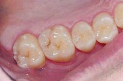
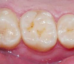
- 31 to 99 — operative care recommended8 (Figures 1 and 2)
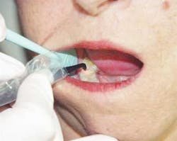
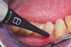
A sterilization cassette contains two site-specific tips that can be placed on the wand: Tip A (Figure 3) is for pits and fissures and Tip B (Figure 4) is for smooth surfaces. When moving Tip A through the fissures or pits, a light pendulum-like motion is used to ensure that the laser beam scans the bottom and sides of the fissure.
Before using the DIAGNOdent, the tooth surface must be free of calculus, plaque, residual prophy paste, and any organic plugs. Air-polishers are an excellent choice to ready the surface for an accurate reading. The surface must also be dry. The device is intended for use on unrestored teeth. Limitations include inaccurate readings around the margins of sealants and existing restorations, due to their innate fluorescence. It is most useful around early, noncavitated lesions.
Studies indicate the DIAGNOdent has high diagnostic sensitivity values of ≥92 percent and is reproducible.9 In a study by Shi, the DIAGNOdent caries-detection method was found to be superior when compared to radiography in discovering early occlusal lesions.10 By extrapolation, the lesions that were once ignored, perhaps "watched" or monitored, are no longer supervised neglect. These lesions can now become confidently diagnosed and proper treatment rendered.
This diagnostic instrument is ideal for remineralization therapy. Each recare interval can welcome and educate patients in their progress against decay, and recall appointments can be tailored to individual caries risk.
Dental professionals have a responsibility to patients to develop a complete caries-management system, including use of early detection devices. Now, clinical decision-making for caries diagnosis must be based on sound scientific research. Dental professionals now have the ability to provide best practices for their patients. This includes early detection of surface changes in enamel, promotion of remineralization therapy in the practice and in home care, and utilization of principles of minimally invasive preparations and restorations.11
References
1 Pitts NB. "Clinical Diagnosis of Dental Caries: A European Perspective." NIH Consensus Development Conference, Diagnosis and Management of Dental Caries Throughout Life, Speaker Presentations. Bethesda, Md.; March 26-28, 2001.
2 Stookey GK. Caries Prevention. J Dent Educ 1998; 62(10):803-811.
3 Black GV. The contact point and its function, considered with reference to dental caries and its treatment. Dent Headlight 1910; 31:135-143.
4 Lussi A. Impact of including or excluding cavitated lesions when evaluating methods for the diagnosis of occlusal caries. Caries Res 1996; 30:389-393.
5 LeGeros RZ. Calcium phosphates in oral biology and medicine. Monogr Oral Sci 1991; 15:1-201.
6 Lussi A, Imwinkelried S, Pitts N, et al. Performance and reproducibility of a laser fluorescence system for the detection of occlusal caries in vitro. Caries Res 1999; 33(4):261-266.
7 Hibst R, Paulus R. Caries detection by red excited fluorescence: Investigations on fluorophores. Caries Res 1999; 33:295.
8 Lussi A, Longbottom C, Braig F, Reich E. Clinical performance of the laser fluorescence system DIAGNOdent for detection of occlusal caries. Caries Res 1999; 33:299.
9 Pereira AC, Verdonschot EH, Huysmans MC. Caries detection methods: Can they aid decision making for invasive sealant treatment? Caries Res 2001; 35:83-89.
10 Shi XQ, Welander U, Angmar-Mansson B. Occlusal caries detection with KaVo DIAGNOdent and radiography: An in vitro comparison. Caries Res 2000; 34:151-158.
11 Trost, L. Today's New Technology: An Improved Caries Removal System. WDJ 2003; 4:48.
Lori Trost, DMD Dr. Trost created the Center for Contemporary Dentistry in Columbia, Ill., in 1989. Her practice is known for being in the technological forefront. She is a member of the ADA and AGD and consults for 3M ESPE's "Council for Innovative Dentistry." You can reach Dr. Trost at [email protected].