It is easy to get wrapped up in the day-to-day routine of doing a crown, filling a cavity, and hopping from one hygiene exam to the next. Our time is important and we have a schedule to maintain, right? While all this is important, the role that we have as health-care providers goes way beyond just fixing teeth — we have a responsibility in the medical community to detect and treat oral cancer.
RELATED |Head, neck, and oral cancer: A general reference for treatment
Why is this important? In the year 2013 alone, more than 42,000 cases of oral cancer will be diagnosed, making it the sixth most common type of cancer (1)— this is approximately 115 cases per day! Our familiarity and frequency to which we are in the oral cavity allows us to most often be the first to identify oral cancer, but in order to do so we have to check for it and look beyond the teeth!
ADDITIONAL READING ...
Oral cancer and the human papillomavirus: Part 1
Oral cancer and the human papillomavirus: Part 2
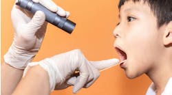
Typically, we see most patients in our care every three to six months. This is two to four times per year we are able to screen for lesions, lumps, bumps, and any atypical pathology that would unknowingly manifest itself to an unaware patient. The routine examination includes the following:
- Update the patient’s health history, which includes medications, surgeries, and blood pressure
- On an appropriate cycle, we can refer to radiographs — bitewing, periapical, and panoramic X-rays
- Assess chief complaints that can be related to oral diseases, which include but are not limited to: halitosis, bad tastes, swellings, too much saliva, dry mouth, delayed tooth eruption, recent occlusal problems, burning sensations, sores, bleeding, and loose teeth
- Complete a head-and-neck exam, palpating for known vs. unknown or suspicious anatomy
- Take into consideration any extraoral lesions that look suspicious or have irregular borders or anatomy
- Evaluate soft tissue in vestibular area, hard/soft palate, dorsum, ventral, and lateral surfaces of the tongue, oropharynx, and lips, paying particular attention to variations in the oral mucosa
If a lesion is found, the following should be considered:
- Note location, shape, size, color, and, if possible, take a photograph
- Record if the lesion is firm, freely movable, or painful to palpation
- Can it be wiped off? Does the patient know about it? Is it unilateral or bilateral?
- Classify the lesion (ulcerative, cystlike, etc.)
- Develop a differential diagnosis and assess risk factors
- The anatomic location of lesions will increase or decrease the likelihood of definitive pathology — i.e., lateral border of the tongue vs. dorsum of tongue
- Follow the protocol established in your office; i.e., two-week follow-up, biopsy, referral, and, if needed, surgery
Case report
Patient X, a 58-year-old male, presented to the office for a new patient exam. Health history included: high blood pressure, he smoked one pack per day (no interest to quit), and had type II diabetes. His last dental visit was approximately one year ago. His chief complaint was that he needed his denture and partial relined. Head/neck exam and all soft tissue within the vestibular and oropharynx were within normal limits. However, a presentation of white corrugated, raised tissue approximately 12x12 mm in size with irregular borders was noted sublingual, toward the patient’s left midline. Patient X stated that he had known of its presence for a short time, but since it was not painful and did not bother him, he did not bother to have it checked. He was referred to an oral surgeon who conducted an incisional biopsy. The diagnosis was mild to moderate dysplasia with areas of carcinoma in situ with areas of likely micro invasion. In light of this finding, patient X was referred to an oncologist for definitive treatment.
Oral cancer screenings can save lives! If detected early, a patient has a 75% survival rate compared to a 25% survival rate if diagnosed late. (1) Not all lesions will be cancerous or pathological in nature, but that is not the point! If we are not seeing beyond the teeth, then we may miss that one opportunity to make a difference!

Reference
1. National Institute of Dental and Craniofacial Research, Oral Cancer,
http://report.nih.gov/NIHfactsheets/ViewFactSheet.aspx?csid=106