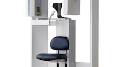
i-CAT, a brand member of the KaVo Kerr Group, is proud to announce the launch of the newest member of the award-winning family of cone-beam 3-D imaging products, the i-CAT FLX MV (medium field-of-view) for general dentists and specialists in periodontics, prosthodontics, endodontics, and oral surgery, so they can place and restore implants and perform oral surgery with greater confidence and lower radiation. The innovative features of the i-CAT FLX MV will deliver greater clarity, ease-of-use, and control for those clinicians who need a medium field-of-view and a range of image sizes to fit a variety of needs.
From ‘scan-to-plan’ to ‘treat,’ i-CAT FLX MV offers these features to seamlessly provide information and control:
- Medium field-of-view captures up to both arches and the temporomandibular joints in 3-D;
- Visual iQuity advanced image technology provides i-CAT’s clearest 3-D and 2-D images;
- Lower dose scan options are available, including QuickScan+;
- Easy-to-use SmartScan Studio touchscreen allows for selection of the appropriate scan for each patient;
- Traditional 2-D panoramic images can be captured with i-PAN; and
- The system can be integrated with CAD/CAM programs.
i-CAT FLX MV offers a balance between image quality and ALARA (As Low As Reasonably Achievable) radiation doses for clinical control and optimized patient care. High-resolution volumetric images provide complete 3-D views to allow for more thorough analyses of bone structure and tooth orientation. QuickScan+ settings allow for full-dentition 3-D imaging at a dose comparable to that which is required for a 2-D panoramic.*
Powerful, clinically driven, comprehensive planning tools streamline workflow and help clinicians move from scanning to consultation and treatment planning in less than one minute. i-CAT FLX MV features Tx Studio 5.3, the latest version of exclusive treatment-planning software with enhanced tools for implants, oral surgery, endodontic procedures, airway analysis, and TMJ treatment. Detailed 3-D images, combined with powerful imaging software, aid in giving dentists the confidence to accurately plan an entire implant treatment from surgical placement of the implant and abutment to the final restoration. Enhance practice efficiency with immediate access to integrated treatment tools for implant planning, as well as access to CAD/CAM applications, including digital models and surgical guides.
3-D scans from i-CAT allow practitioners to perform more advanced procedures with greater predictability. i-CAT’s open software architecture seamlessly integrates with orthodontic systems, CAD/CAM programs, imaging software, and practice management programs, expanding the practice’s capabilities.
“With the launch of the i-CAT FLX MV, dental professionals can invest in a medium field-of-view that captures the anatomical area specific to their treatment focus,” states Rick Matty, Director of Marketing for i-CAT. “As a result, we are serving more dentists by providing i-CAT quality in a model that fits most every practice.”
i-CAT has evolved into the most trusted 3-D imaging brand, widely regarded as the industry standard in cone-beam technology. i-CAT solutions have been installed in more than 5,000 sites around the world. To help dentists make the most of i-CAT, i-CAT offers highly specialized service and support through the i-CAT Network and continuing education through i-CAT University, the only entity of its kind dedicated to helping dentists and specialists. Learn more about education and the i-CAT FLX family of products at i-cat.com.
*Data on file; Indications for Use at i-cat.com/ifu.


