This is a terrible title for an article about endodontic files, and to an animal lover it may even seem inappropriate, but it is an expression we have all heard referring to the fact that in many situations there are multiple alternatives to achieve the same end result. This idiom, however objectionable, has become more relevant to the discussion of endodontic files in the last several years due to the explosion of the various file systems that have flooded the marketplace. With all of the confusion that has developed around what might be the best file system, it has become increasingly more difficult for the practitioner to make an informed choice about which endodontic files can achieve the best clinical outcomes.
ALSO BY DR. LANDWEHR |Electronic determination of working length: more accurate than radiographs?
Because of the incredible anatomical complexity within the root canal system and the highly variable size, shape, and length of roots, the number of file systems that have been introduced should be considered a major advancement in endodontic treatment. With all of the file choices available to the clinician, it is now possible to create a more customized final shape in the root canal, which can be determined by the preoperative anatomy.
ALSO BY DR. LANDWEHR | Endodontic case study: referred pain leads to diagnostic uncertainty
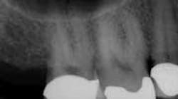
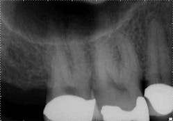
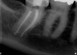
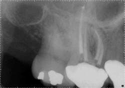
For example, in canal anatomy that appears to be fairly straight on preoperative radiographs in roots with traditional taper, a single file system such as WaveOne(Dentsply Tulsa Dental Specialties) would be an excellent choice to achieve cleanliness and shape (Figs. 1 and 2). The single-file reciprocating system would also have the added benefits of simplicity and efficiency.
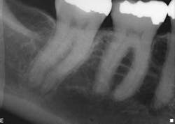
However, when the anatomy is the most complicated with multiple curves or the smallest canals, a different file system might be a better choice (Figs. 3 and 4). Although almost all root canals can be treated with WaveOne, in certain anatomies the root canal procedure is actually easier and more efficient when utilizing a file system that has more tip sizes and taper choices and ultimately requires more files. Vortex Blue (Dentsply Tulsa Dental Specialties), with its incredible resistance to cyclic fatigue and reduced shape memory, is an ideal file choice in this clinical situation. Vortex Blue files have tip sizes ranging from ISO 15-50 and tapers of 0.04 and 0.06. This results in great flexibility to treat the most complicated canal anatomies — flexibility not only in the file itself, but also in the size and taper choices that are available. However, to appreciate the metallurgical advantages of the Vortex Blue files, it may be necessary to spin four or as many as six files per canal.
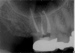
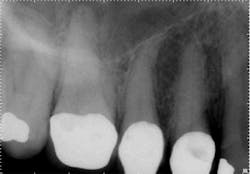
When the anatomy is more complicated than can be predictably treated with WaveOne, but not so irregular as to require all of the files of Vortex Blue, is there another option? It is in these clinical situations that ProTaper Next® (Dentsply Tulsa Dental Specialties) becomes the best file system (Figs. 5 and 6). The rectangular but off-center cross-section creates the illusion that the canal is slightly larger than it is because the alternating contact points of the file prevent the feeling of the file getting locked into the canal space. This added space along the file results in additional room for debris removal, and in spite of this design, there is no appreciable loss of cutting efficiency. When using ProTaper Next files to create the final root canal shape, two or three instruments will be used.
In these three cases, different instrumentation systems were used to create the final shape of the root canal system. In spite of differences in the number of files used, the file design and file movement, the end results look very similar. Although potentially confusing to clinicians, the advances in metallurgy and file design allow for safer and more efficient cleaning and shaping of the root canal system than ever before. However, despite these advances, the success rates of endodontic treatment have stagnated over the last several decades, indicating there is more to clinical success than a dense, white line on a radiograph. Canal shaping is important, but it is paramount that an effective irrigation protocol is incorporated into the endodontic procedure to safely and effectively eliminate all possible bacteria.Fig. 1: Although access is usually an issue when treating second molars, the shape of the roots and good chamber size suggest WaveOne would be an ideal file system for tooth No. 2.
Fig. 2: Following establishment of a glide path and removing the dentin triangle, a single WaveOne primary file was used to create the final shape. This shape would allow for deep and safe delivery of irrigants and maximum disruption of biofilm.
Fig. 3: Preoperative anatomy of tooth No. 31 shows a double curve in the mesial root canal. The size, length and taper of the root suggest a file system with multiple tip and taper choices would be most suitable.
Fig. 4: A glide path was established with a 15/0.04 Vortex Blue rotary file in conjunction with size 10 hand files. Vortex Blue files were then used in sequence until a final shape of 30/0.04 was achieved in the mesial root and 30/0.06 in the distal root.
Fig. 5: With a crown in place, it can be difficult to determine the size of the pulp chamber. However, at the level of the roots, the canals in tooth No. 3 look to have calcified compared to the neighboring teeth.
Fig. 6: After locating the canals and following the glide path to the root ends with hand files, two ProTaper Next files were used in each root canal to create the final shape.
