Mary Sue Stonisch, DDS, FAGD, AAACD, DICOI, shares her approach to dental photography, facially generated smiles, and case acceptance.
The way we gather data in dentistry—from simple digital photography to 3-D imaging—has transformed how we make decisions. What has not changed is where the teeth fit in the face and smile. We need to be able to communicate an ideal smile to a patient before he or she has committed to comprehensive treatment, and our communication with the patient needs to be reasonable in cost, easy to expedite, and personalized. For these reasons, using advanced technology may not be the best choice for every patient.
Diagnostic wax-ups, mock-ups, and digital smile design all require time and money to achieve, and fee-for-service dentists are now being challenged and squeezed by corporate dentistry. We need to be proficient and economical in gathering records for treatment planning so we don't price ourselves out of the market.
At "hello," most of us are already assessing the horizontal plane of occlusion and visual asymmetries. But what happens once the patient leaves the office? Unless we have captured appropriate data through photographs and model work, that visual assessment will be useless. So where do we begin? After getting a thorough medical and dental history, it’s as simple as one-two-three. Just three procedures can simplify your dentistry and increase your profitability: (1) documentation through photography, (2) establishing a new tooth length and plane of occlusion, and (3) model work and jaw measurements.
Step No. 1: Document through photography
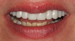
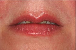
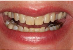
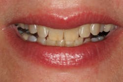
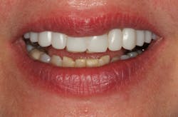
Quality photographs are essential. In my practice, four simple dental views are mandatory: a smile, a retracted view, a view of the mouth in repose, and a Duchenne smile. Especially critical are the repose view and the Duchenne smile shot. These two views allow us to be efficient in treatment planning. Have you ever completed a smile rehabilitation and missed the mark on the tooth length? The patient’s tooth edge is not even visible with the lip at rest. Quite unfortunate, don’t you think? Have you ever completed a smile makeover and wished you had lifted the gumline higher? The patient could have smiled bigger following rehabilitation, due to increased confidence and reduced gumminess! Both of these pictures are essential in assessing the goals of smile enhancement.
A Duchenne smile
Step No. 2: Establish a new tooth length and plane of occlusion
Establish a desired maxillary incisal edge position for the patient's two front teeth and whether a change in the horizontal plane of occlusion is necessary. Gathering this information before the patient leaves the smile consultation can be the first step in a successful case outcome financially and esthetically. This is an essential second step before a facially generated treatment plan can begin.
Step No. 3: Take jaw measurements and develop models
Model work is essential to send a patient home and work on the patient's case on the lab bench. Assuming there are no bite issues, simple models and a few measurements can facilitate transferring this information to the articulator.
Now the fun begins! Smiles can be generated in wax easily. Information can be shared with the patient to verify the proposed smile design and confirm the incisal edge position, plane of occlusion, and functional concepts for a new oral rehabilitation.
If we are to keep up with the competition, these simple principles are the ticket. There is no need for sophisticated imaging, digitalization, or computer skills. Simply apply the basics along with this inexpensive Smile-Now template and voila! The end results can be a gorgeous smile on a budget.
