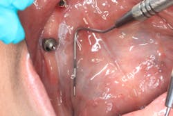
Dr. Stacey Simmons describes the oral pathology case of a 60-year-old patient who presents for a limited exam. Chief complaint: She noticed some white tissue under her lower implant prosthesis. The patient is concerned because a similar lesion was removed and diagnosed as precancerous five years ago when her implants were placed.
Editor's note: This article first appeared in DE's Breakthrough Clinical with Stacey Simmons, DDS. Find out more about the clinical specialties newsletter created just for dentists, and subscribe here.
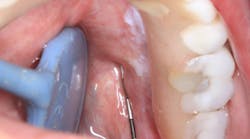
Figure 1
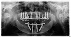
PRESENTATION: A 60-year-old female with a noncontributory health history presents for a limited exam.
CHIEF COMPLAINT: The patient noticed some white tissue under her lower implant prosthesis (figure 1). The patient reported no pain in the area but was concerned about the white tissue being something pathological. Apparently, when she had her implants placed five years prior, a lesion of similar nature was removed and diagnosed as precancerous.
THREE MONTHS EARLIER: The patient had been seen three months earlier for a recall exam and lower prosthesis removal and cleaning. Findings at the soft-tissue exam were within normal limits.
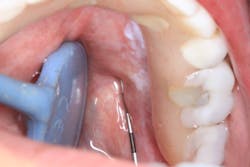
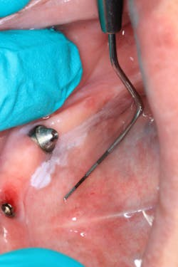
CLINICAL EXAM: Clinical assessment reveals a white, corrugated lesion lingual to the acrylic of the fixed hybrid prosthesis (figure 2). The lesion measures 5x24 mm and is not able to be scraped off or removed. It is not painful or symptomatic, and there are no swellings noted in the sublingual or submandibular lymph node areas (figures 3 and 4).
Figure 2
Figure 3
Figure 4
What are your differentials and recommended course of treatment modalities?
Send your answers to [email protected] or join our Facebook group to discuss this oral pathology case and more. Next month, we will discuss the final diagnosis and recommended treatment for this case.
Do you have an interesting oral pathology case you would like to share with Breakthrough’s readers? If so, submit a clinical radiograph or high-resolution photograph, a patient history, diagnosis, and treatment rendered to: [email protected]. We will let you know if we select your case.
For more oral pathology articles, click here.
Editor's note: This article first appeared inDE's Breakthrough Clinical with Stacey Simmons, DDS. Find out more about the clinical specialties newsletter created just for dentists, andsubscribe here.
