Delivering the message -- facilitating communications with cone beam
By Walter Chitwood, Jr, DDSIt has been said that “knowledge is power.” Keeping current with technological advancements and new treatment options is a time-consuming, but very necessary endeavor. However, just collecting the knowledge is not enough. To grow a successful and profitable practice, one also must be able to effectively communicate these new treatment methods to patients and colleagues. Digital radiography, specifically through 3-D imaging, is one way that I have improved my communication skills.While I build communication with carefully chosen words and experience, cone beam imaging improves patient understanding even more because of its 3-D capability. Most patients are not trained in reading X-rays and do not comprehend the clinical signs on those X-rays that concern dentists the most. Cone beam’s 3-dimensional view of the dentition can be enlarged, rotated, or cross-sectioned at any angle, so patients can see what we see — undistorted images of bone structure deterioration and nerve canals that encourage compliance with our treatment plan. When patients can see their own anatomy in 3-D on a large computer monitor, they become more confident in the diagnosis.
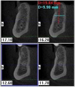
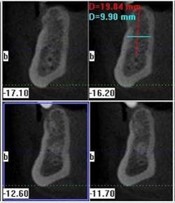
The power of cross-sections and precise measurementsCommunicating with conviction is also a key to treatment acceptance. The detail provided by cone beam images takes the guesswork out of implant planning. Dentists without 3-D images must account for the scenarios that sometimes pop up as a result of conditions that we cannot discern from less detailed imaging options. They must inform the patient that the treatment plan may change, depending on what is discovered during the first surgical session. Discussing these “what-if’s” with the patient leads to doubt and procrastination. A cone beam scan offers a view of the details that were previously only visible during surgery. When patients comprehend their condition, they are more eager to begin treatment quickly. Cone beam helps there too. Since my GXCB-500™ unit fits right in our office, the patient can go from imaging to diagnosis to treatment in less time. With in-house 3-D capability, my communication for implant planning and placement has also improved with referring dentists who want to stay current with their patient’s progress. To facilitate this, I print out a screenshot of the patient’s dentition and e-mail it to the referring dentist, or send a CD with the imaging software and the scans. If the referring dentist wants to discuss the situation, all the facts are on both of our computers in 3-D, but often the clarity of the images removes any doubts. These dentists are happier because they don’t want their patients to have any surprises during treatment that may affect them later either.
Easily understood treatment plansStaying aware of the latest innovations in our field by attending conferences, taking CE classes and courses (such as the ones my 3-D company offers at the 3-D Imaging Institute in Raleigh, N.C.), and reading dental journals are just a few ways that I gain insight into groundbreaking technologies to better serve my patients. While I amass all of the information that I can to add depth to my practice, my cone beam scans truly add dimension to my professional relationships. The knowledge and communication opportunities provided by CBCT benefit patients, referring dentists and specialists, and result in a more productive practice.
Dr. Walter Chitwood is a 1978 graduate of Middle Tennessee State University and the University of Tennessee College of Dentistry in 1982. His postgraduate training includes implant externships at the Center for Oral and Maxillofacial Rehabilitation in Clearwater, Fla., and the Misch Implant Institute in Dearborn, Mich. Dr. Chitwood is a diplomate of the American Board of Oral Implantology/Implant Dentistry, diplomate of the International Congress of Oral Implantologists, and honored fellow of the American Academy of Implant Dentistry. He is a fellow of several organizations including the American Society of Osseointegration, the Academy for Implants and Transplants, and the Academy of Dentistry International. In addition to his private dental practice, he holds board of director positions for several dental organizations including the American Board of Implantology/Implant Dentistry and the Tennessee Academy of General Dentistry. He is also an educator. Dr. Chitwood’s office is an off-campus site of dental education for the Tennessee Technology Centers, the source of education for certified dental assistants in his state. Over the years he has held several teaching positions including assistant clinical professor at the Vanderbilt School of Medicine, Department of Hospital Medicine, adjunct professor at Tennessee State University, and director for the Professional Resource Education Center also located in Murfreesboro, Tenn.


