The RF Bleeding Index: A better way to detect active interproximal gum disease?
This article is to introduce a possible better method of evaluating the intactness or health of the interproximal sulcular epithelium, the RF Bleeding Index technique. This technique also helps to determine when these tissues need additional treatments to regain their health.
Intact sulcular epithelium may reduce the invasion of sulcular microbes into our systemic circulation. Researchers continue to emphasize the probable adverse effect on our systemic health posed by these invasive oral microbes.
This article will not delve into the deleterious effects of these invasive microbes, but discuss a technique for dentists, hygienists, and patients to simply and quickly assess the interproximal sulcular epithelium’s need for treatment, and help monitor the ongoing treatments effectiveness in healing these tissues.
The reasoning
There are many fine assessing indexes, as discussed in Sven Paulsen’s paper. (1) All these indexes raise concerns about the amount of time needed to perform multiple insertions of the periodontal probe and then the charting of each measurement.
Periodontal probe depth may not be an indicator of active disease, unless active bleeding results following this probing. Sulcular epithelial ulcerations may be like small spots of rust on an otherwise intact rain gutter, or they can be broad swaths of pervasive rust (ulcers) sweeping across all surfaces. Using any of the standard periodontal probes, you may or may not hit one of the spotty ulcerations, and show minimal to no bleeding. This might give the evaluator a false sense of gingival health. I felt I needed a more comprehensive attempt to find ulcerated sulcular walls.
Over time, I developed an idea outlined in “The Swedish Wonder: A better way to control gum disease and decay and cover floss’s shortcomings.” The concept was published along with videos in DentistryIQ in January 2017.
Some of the most damaging of the oral microbes grow somewhat undisturbed below the reach of dental floss. This undisturbed biomass may in time convert to an anaerobic biomass with tissue damage rates not easily controlled by our oxygen-dependent immune cells. The Swedish Wonder technique allows better control of these microbes by ideally removing their food and disrupting colony development.
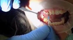
All interdental pics that are safe to use in this technique I call “Swedish Wonders” or “Swedes.” Gum’s Soft-Picks for Wider Spaces (blue tip) or Original (green tip) are Swedes of choice and are placed to the depth of every interproximal sulcus. This is done ideally following any main meals or snacks by allowing the flexible non-sharp tip to ride the sulcus floor from facial to lingual displacing food and biomass. The reader may see this technique by viewing the three videos below.
A part of developing a simplified test to detect ulcerated sulcular walls is having an equally simple treatment approach that can be modified to match apparent severity of the infection. The RF Bleeding Index is performed with the exact same motion of the Swede as shown in the videos at the bottom of this article.
In other words, you insert the pick on the mesial or distal of each tooth on the facial aspect vertically down to the sulcular floor, and ride the floor from facial to lingual while keeping the tip on the sulcus floor. Then you smoothly withdraw the pick and chart the degree of bleeding coming from the point of withdrawal as shown in the video. In my experience of over 30 years following patients who used the Swede correctly, there was no apical migration of the epithelial attachment (attachment loss) or increase in pocket depth. It is the destructive action of the microbes that is the root cause of attachment loss. Therefore it follows if you remove the microbes' food and disrupt their maturation and growth, you stabilize interproximal periodontal health.
Because of the depth the interdental brush achieves and its size and soft projections, most sulcular wall ulcerations have their soft clotted ulcerous scabs dislodged. The resulting bleeding is what is charted.
I base my selection of treatment regimens by the amount of bleeding on withdrawal of the Swede:
For patients with interproximal charted pocket depths from 2 to 4 mm:
- No bleeding on withdrawal: No additional home care tasks needed at this time
- 1-2 mm of blood above gingiva: Swede one to two times per day to depth of the pocket
- Half of interproximal space filled: Swede after each main meal to the depth of the pocket
- Full interproximal space filled: Swede to the depth of the pocket after eating anything
- In five to 10 days, most bleeding will have been reduced in those patients sticking to the regimen, and then some may try a lower frequency of use. If bleeding or odor returns, then you must bring up the frequency of the Swede use again.
For patients of interproximal probings of 4 to 8 mm:
- Swede should be used to the depth of the pocket with the sweep as shown in the video with each consumption of food
Until one company develops a durable probe that a dentist or hygienist may use during patient examinations for the RF Index, the Gum Soft-Picks (blue and green tips) are the instruments of choice for RF Index evaluation. The Original (green tip) is to be used for very tight interproximal spaces. Never use any picks with wire-incased bristles or ones with sharp ends. Please advise patients that the initial placement of Swede may be uncomfortable as it will contact ulcerated tissue, but most discomfort and bleeding will be gone in two to 15 days. It may never bleed again for those that stay with the daily regime. Caution patients against more than one to two insertions for each interproximal space following each meal, as some people may develop inflammation from being too zealous with many back and forth motions.
Conclusion
Having a quick and simple way to assess the degree of interproximal gingival sulcular wall ulceration or disease makes for better patient compliance, especially when the patient experiences a cessation of bleeding and lowering of discomfort from the Swede regimen.
One of the greatest benefits for me, as it hopefully will be for all the dentists or hygienists who take up the promotion of the Swede, is to put you more in control of adverse patient demands such as, “ I am still bleeding and it hurts,” or “You just put this crown in, and now it needs redoing!?” My query to these expressions of dissatisfaction and my opening to every exam of current patients is usually, “Have you been faithful with your Swede use?” The reason you can feel less stress in these situations is that proper use of the Swede works to reduce failure, bleeding, and soreness consistently.
The RF Bleeding Index is only a small additional step in the detection of interproximal gum disease and the Swedish Wonder is just an addition to brushing and flossing and any home care steps your patients are currently using. They do not replace regular dental exams and treatment.
Very best of success for you with this concept. It has been a great help to my patients for many years.
Reference1. Poulson S. Epidemiology and indices of gingival and periodontal disease. Pediat Dent. 1981;3.
William Fell, DDS, is a member of the 1968 first graduating class of UCLA dental school. He was involved with research projects on electronic patient interaction, patient data, instruction and student abilities to review all lectures after the fact at electronic libraries. He also did research to improve casting accuracy at UCLA. He has dental and nondental patents. His article, "One visit composite and amalgam bonding for strong aesthetic posterior restorations,” has been published in JADA.
About the Author
William Fell, DDS
William Fell, DDS, is a member of the 1968 first graduating class of UCLA dental school. He was involved with research projects on electronic patient interaction, patient data, instruction and student abilities to review all lectures after the fact at electronic libraries. He also did research to improve casting accuracy at UCLA. He has dental and nondental patents. His article, "One visit composite and amalgam bonding for strong aesthetic posterior restorations,” has been published in JADA.

