Sealants ... what a great idea!
By Sheri B. Doniger, DDS
Sealants have been around, in some form or another, for almost 40 years. Methods to eradicate occlusal decay have been in place for more than 100 years prior to the evolution of dental sealants. GV Black's "extension for prevention" involved removing healthy dentin and enamel areas beyond the apparent decay. Prophylactic odontomy, developed by Hyatt, involved eradicating pits and fissures then packing them with amalgam. Bodecker recommended enameloplasties, so fissures were packed with cement. It was the right idea, but they weren't retained. Sealants first came to the forefront by the adhesive resin research of Dr. Bunacoure in 1965, and were finally put into practice by Simonsen and Stallard in 1967.
According the CDC Oral Health 2000 Facts and Figures, tooth decay still remains one of the most common childhood diseases. Eighteen percent of 2- to 4-year-olds experience tooth decay, and 16 percent still have untreated lesions. By the age of 17, 78 percent of children will have experienced dental disease, with 7 percent losing at least one permanent tooth. Occlusal surfaces account for 12.5 percent of all tooth surfaces, but will experience more than 50 percent of all decay. Pits and fissure caries account for 88 percent of total caries in children. Fluoride is not as effective in pits and fissures as it is on smooth surfaces. Tooth susceptibility to molars remains the highest. Our main goal is to prevent decay and maintain the health of the patient's dentition throughout life. Dental sealants offer a safe and effective method to prevent dental decay.
Sealants have a definite place in the preventive practice. "Sealants are 100 percent effective in preventing pit and fissure caries if they are completely retained," a recent article stated. With proper placement techniques, sealants in place for up to 15 years are common. Sealants need to be repaired and restored to continue to be beneficial. Approximately 2 to 4 percent of all sealants placed need some form of repair over the lifetime of the tooth. This repair usually occurs during the first year. Sealants placed on the buccal or lingual surfaces of the tooth have the highest rate of failure. These teeth may need filled materials or adjunct preparations to allow for increased surface area retention. It is estimated that 10 percent of occlusal sealants are lost during the year, but more than 30 percent of buccal/lingual sealants are lost.
The problem is that sealants are still incredibly underutilized. There are several reasons for underutilization. Although sealants have proven effective over the past 15-plus years of study, insurance companies still do not pay for dental sealants on a routine basis. They would rather pay for permanent restorative care, such as amalgams or composites that cut into the tooth. The fee for a dental sealant is approximately one-third the cost of a permanent restoration. It is inconceivable why insurance companies would prefer to cover surgical intervention of a tooth rather than a preventive intervention.
Historically, underutilization could be blamed on lack of knowledge by clinicians. Currently, with all of the good data available, especially concerning long-term success rates, it is more the lack of knowledge by patients. We need to educate our patients, their employers, and insurance companies about the value of a mechanically bonded surface protecting sealant over a restoration that will most likely need larger replacement over the history of the tooth. Some patients will rely on insurance coverage — "If the insurance company doesn't pay for it, I don't want it." A patient's refusal of treatment on the basis of "lack of insurance coverage" is an excuse that may be rectified by proper education. The overall cost savings of a caries-free tooth and the benefit of an intact dentition need to be stressed.
The entire dental team needs to be focused on the preventive nature of the sealant. Every team member needs to be knowledgeable on the procedure and the benefits. The business coordinator should have materials available for discussion when the parents visit with the children. In-office literature depicting sealant placement and rationale would be beneficial to patient education. The assistants can use demonstration models, prepared with a sealant placed on a sterilized extracted tooth. Procedures can be demonstrated to both child and adult to show the relative ease of placement. Some offices offer audio-video or CDs with patient education for viewing in the reception area. Even though this is a dental procedure where no anesthetic is utilized, patient fears need to be overcome. The more the patients understand the concept of prevention, the more likely they will accept the treatment plan.
Since caries is a multifactor disease process (the susceptible host surface, available bacteria, and sucrose supply), we need to assess the risk of the tooth for the development of the disease. Caries risk factors include poor oral hygiene, cariogenic diet, familial history, low fluoride intake, and history of dentinal caries. The patient's history of preventive care, current orthodontia, medical issues such as xerostomia, and pit and fissure anatomy are also considered. Teeth should be assessed individually to determine their risk. Caries is also now considered to be a cyclic process that involves mineralization and demineralization of the tooth surface. The saliva, antimicrobials, calcium additives, and fluoride will mineralize while the oral bacteria and sugar will attack the surface and demineralize. Early detection is very important in preventing the irreversible carious demineralization.
If teeth are caries-free, they can be sealed or monitored if the tooth or individual is not at a high risk for developing decay. Individual dentists have differing views in practice. Sealants should be placed on all susceptible teeth, especially molars, as soon after eruption as possible. Teeth should have no operculum or tissue flap present, as this would prohibit complete coverage of the occlusal table with the resin. The teeth should exhibit susceptible morphology with deep grooves and fissures present on teeth that we wish to seal. A tooth with a flat morphology would be less in need of a sealant than a tooth with a multiple, deep-grooved pattern. Radiographs should be taken prior to sealant placement to assess the lack of interproximal decay. Studies have shown that sealing in incipient decay will block off the nutrient source from the carious lesion, rendering it inactive rather than active. Frank occlusal decay should not be sealed, nor should primary teeth that are close to exfoliation.
Contra indications to sealants do occur, including a patient's behavior management preventing proper sealant placement, inability to isolate and maintain a dry field, semi-erupted teeth, presence of decay, and an allergy to methacrylate.
Resin sealants, usually composed of bisphenol a-glycidyl methacrylate (BIS-GMA), form a mechanical retention to the tooth enamel. This occurs when the acid etch (usually a 30 to 37 percent phosphoric acid gel or solution) is applied to the tooth, removing part of the organic matrix of the enamel rod. Pores are made in the enamel, thereby increasing the available surface area of the tooth, then filled with resin (resin tags). These tags mechanically hold the resin to the tooth. The acid etch will also remove pellicle, plaque, and debris from the tooth surface. This etching procedure is also called conditioning. Glass ionomer-based sealant materials utilize a chemical bond to the tooth structure.
Sealants may be either chemically or auto polymerized. Chemical or self-cured sealant has two components — a catalyst and a base — which are mixed together to begin the chemical reaction. With this type of product, the setting time — usually a 60- to 90-second working time window — is determined by the manufacturer. The material is mixed, then placed on the tooth. A small amount will remain in the mixing dish, which can be "tested" for sealant hardness, prior to using the explorer on the freshly sealed tooth surface. Auto or light polymerization occurs with either a high-intensity visible light or a laser. Historically, UV light was utilized. These types of sealants have a more command set, allowing the operator reasonable time to place. The sealant is placed on the tooth, exposed to light, and is set, usually within 20 seconds.
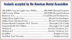
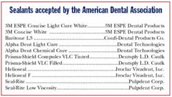
Sealants may be also classified as unfilled or filled. Unfilled resins are BIS-GMA only. They have a high ability to flow, but may not be easily retained on vertical or buccal surfaces. Unfilled resins are either clear or colored. Filled sealants contain glass, quartz rods, or silica in varying percentages, ranging from 40 to 60 percent. The higher the amount of filler, the more abrasion-resistant is the sealant. Filled sealants are usually light-cured, and can be either opaque or tinted. One brand of sealant changes color after it is applied to clear. Another will show a color when exposed to light-cure during recall appointments, demonstrating the integrity of the sealant. Cyanoacrylates are also utilized as sealant material. Some sealants have a fluoride-releasing component, which is either a fluoride salt or a fluorosilicate glass. This fluoride will assist in remineralization. Resin-reinforced glass ionomer sealants are also available which chemically bond to the tooth and have a high fluoride-releasing ability.
Success of the sealant treatment depends on the placement technique. A dry field is paramount to retention. Moisture contamination is the biggest reason for sealant failure. Bubbles in the resin will cause a weakness of the sealant coat, and incomplete groove coverage will also lead to sealant failures and possible decay. It is the responsibility of the clinician to check for the integrity of the sealant each preventive visit. Sealants should be repaired if coverage is compromised.
The initial step of sealant placement is the determination of its need on a particular tooth surface. Several methods of caries detection are available. The standard visual examination-tactile explorer-radiograph technique is the most common being utilized. Caries detection dyes, though available, may not offer the precise diagnostic ability as an explorer and tactile sensitivity. Some dyes will irreversibly stain the enamel.
Newer technologies are now emerging to detect early demineralization and caries. Laser fluorescence uses a single wavelength of light (655 nM) to find porphyrines in carious tissue. DIAGNODent (Kavo, pictured at right) is the tool that is directed to the occlusal surface to detect demineralization. Fiber optic transillumination and digital imaging fiber optic transillumination (DIFOTI) utilizes an intensified light to be focused on the tooth. It is capable of determining both occlusal and interproximal demineralization. Quantitative light-induced fluorescence (QLF) utilizes fluorescence to determine demineralization in occlusal, buccal, and lingual surfaces.
Depending on the depth of the fissure and the need to remove enamel decay, the dentist has several options. Air abrasion and microdentistry to open fissures and remove enamel decay may be performed prior to sealant application. Both high- and low-speed tooth preparation may be utilized. The assistant may be able to restore the tooth after these surgical procedures are performed, depending on the particular state expanded function rules.
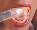
Retention of sealants depends on moisture control. Several moisture-control methods are available. Isolation with rubber dam, cotton rolls, and absorbent pads are the most common. A new innovation, Isolite (pictured at right), is a remarkable product that will isolate two quadrants, retract the tongue, prevent closure of the mouth, evacuate fluids, and light up the entire oral cavity, allowing the clinician to visualize and seal with the appropriate dry field. For the practitioner placing sealants alone, this is an essential addition to the office armamentarium.
Various methods of cleaning the tooth have been studied. Explorer, toothbrush, pumice, hydrogen peroxide, and air slurry polishing are utilized. If air slurry polishing is utilized, a dual etching or conditioning procedure must be applied to neutralize the sodium bicarbonate. The use of fluoride prophy paste will not affect the bonding of the sealant to the tooth, so sealants may be performed at the same visit as a prophylaxis.
Some studies are suggesting the use of a dentin-bonding agent prior to sealant placement, but this adds another step to the process. After cleaning, tooth conditioning and washing, two applications of a third- or fourth-generation dentin bonding agent are placed after a brief drying period. The sealant is then placed. The enamel rods in primary teeth do not form a perpendicular angle to the surface, as do permanent teeth. The monomer in the bonding agents penetrates the enamel rods more effectively, and some believe this will improve retention.
Following sealant placement, the tooth should be evaluated for sealant integrity and coverage. After wiping off the air-inhibited layer of nonpolymerized resin, the use of an explorer at the margins is recommended. The sealant should be visually evaluated for bubbles or voids in addition to an evaluation for lack of occlusal interferences. The use of dental floss is recommended to ensure open contacts, especially if a clear sealant is utilized. Recall evaluation of sealants should be made at preventive maintenance appointments.
Although fluoride varnish is an adjunct tool for desensitization and alternative fluoride delivery, success in application of a varnish in lieu of a dental sealant has not been documented. The varnish adheres to the fissure and allows for fluoride release over a period of time. The varnish applications need to be repeated and their efficacy in pits and fissures are not as effective as the placement of a sealant.
Glass ionomer cement is now being utilized as a sealant. The ability to place glass ionomer in a wet environment is a definite plus with a newly erupted tooth, even with operculum coverage. Studies have shown that if the glass ionomer is not completely retained in the entire sealed area, the amount left in the pits and fissures still releases fluoride that is effective in reducing caries. The glass ionomer is chemically bonded to the enamel and dentin and has been proven to be cariostatic.
Triage, a Fuji product, has a 24-month fluoride release, works in a wet field, and is self-bonding, allowing for fewer steps in the process of application. The product has a low viscosity and can be spread over the pits and fissures. This may be advantageous in certain behavior management cases or less-than-perfect tooth surface placement. It allows for coverage and fluoride protection during a highly susceptible period of early tooth eruption.
Another unique new product is Embrace from Pulpdent. It is less technique sensitive as compared to traditional sealants. The sealant is a hydrophilic resin acid-integrating network, which forms a chemical bond to the tooth and is placed in a wet environment. Since it is a wet bond, it will be effective in the presence of water. In previous sealant materials, saliva contamination would be a reason to begin a re-etch process. With this product, a small amount of saliva would allow for the wet environment necessary for the activation of the sealant.
After etching, the tooth is rinsed but not dried. This sealant material mixes with the water and becomes part of it. The resin becomes less viscous when placed and is drawn down into the pits and fissures. The sealant is tooth-colored with a fluoride release. Since the material integrates with the tooth, the margins are not discernable. This makes the material less resistant to marginal wear and eliminates microleakage. Studies have shown 98 percent retention after one year.
The Healthy People 2010 initiative objective 21-8 states, "increase the proportion of children who have received dental sealants on their molar teeth." Their goal is to have 70 percent of all children, aged 8 to 14, have at least one molar sealant by 2010. The initiative fell short in the Healthy People 2000. To achieve this goal, there is a need for further patient education about the benefits of sealants.
With the cost effectiveness of sealant placement by auxiliaries, more sealants should be placed by dental assistants to increase utilization. Many states offer sealant placement as an expanded function for an auxillary. Utilization of an assistant in sealant placement serves a dual service — to the patient for the marvelous preventive care option and to the practice for the cost-effectiveness. If sealants are charged out at a rate of $40 to $60 per tooth, and four teeth can be easily sealed in 30 minutes, this will give a minimum hourly production rate of $320 to $480.
Sealants are one of the best preventive measures we can offer our patients. With proper placement, a sealant has proven longevity and will protect the occlusal surface of the tooth from decay. These procedures are esthetic, non-invasive, and performed without anesthetic. Simply put, this is a cost-effective method of prevention for our patients.
Sealants need to be maintained throughout the patient's life for a caries-free tooth for a lifetime. Along with proper dental care, fluoride treatment, patient compliance for oral hygiene, and sealant placement, we have the tools to help our patients keep their teeth for a lifetime. The future of dental health is now. Let's make sealants a bigger part of our office's preventive goal.
Editor's Note: The author wishes to thank Mr. Jeff Gartman and the American Dental Association Library staff for their ever-gracious assistance. References for this article are available upon request.
Dr. Sheri B. Doniger practices in Lincolnwood, Ill. She graduated from the University of Illinois College of Dentistry in 1983 and obtained her bachelor's degree in dental hygiene from Loyola University of Chicago in 1976. She can be reached at (847) 677-1101 or [email protected].
Risk factors, as modified from the American Dental Association Council on Access, Prevention and International Relations
Children
Low Risk
No new or incipient caries in the past year
Good oral hygiene
Regular dental visits
Moderate Risk
One new, incipient or recurrent caries in the past year
Deep or non-coalesced pits and fissures
High familial caries experience
History of pits and fissure caries
Early childhood caries
Frequent sugar exposures
Decreased salivary flow
Compromised oral hygiene
Irregular dental visits
Inadequate fluoride exposure
Proximal radiolucency
Orthodontic treatment
High Risk
Two or more new, incipient, or recurrent carious lesions in the past year
Deep or non-coalesced pits and fissures
Siblings or parents with high caries rate
History of pit and fissure caries
Early childhood caries
Frequent sugar exposures
Decreased salivary flow
Compromised oral hygiene
Proximal radiolucency
Adults
Low Risk
No new or incipient caries
No more than one recurrent lesion during the past three years
Good oral hygiene
Regular dental visits
Moderate Risk
One to two new, incipient or recurrent caries in the past year
History of numerous or severe caries
Deep or non-coalesced pits and fissures
Frequent sugar exposures
Decreased salivary flow
Irregular dental visits
Inadequate fluoride exposure
Exposed root surfaces
Fair oral hygiene
High Risk
Three or more carious lesions in the past three years
History of numerous or severe caries
Deep or non-coalesced pits and fissures
Frequent sugar exposures
Decreased salivary flow
Irregular dental visits
Inadequate fluoride exposure
Compromised oral hygiene
Sealant placement protocol
- Caries detection
- Isolation
- Tooth cleaning
- Rinse for 30 seconds
- Tooth conditioning for 60 seconds
- Rinse for 60 seconds
- Re-condition if air slurry polishing is utilized
- Air dry tooth until chalky, frosty appearance
- Sealant application
- Curing (if applicable)
- Evaluate placement
- Check occlusion
- Post operative instructions to patient
- Re-evaluation of sealant at preventive appointments


