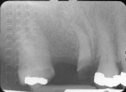
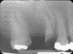
Dr. Antenucci giving a lecture on 3-DThese are challenging times then for both dentists and patients alike who are sensitive about radiation exposure. But according to Gene Antenucci, DDS, of Huntington, N.Y., and a consultant to
Planmeca USA, there are features in dental imaging units that can reduce radiation exposure.Dr. Antenucci encourages dentists to look for 3-D imaging units with built-in safety measures that focus on using the least amount of radiation to achieve the best results possible. This commitment to “As Low As Reasonably Achievable” — or the ALARA radiation principle — keeps both the dentist’s and patient’s safety at a priority. Progressive, safety-conscious manufacturers are increasingly designing their imaging units with a multitude of different volume selections, which allow the dentist to radiate only the area of clinical need. That, combined with the technology to change the radiation amounts more precisely, helps keep radiation dosages as low as possible for patients. Dr. Antenucci encourages his colleagues to look for units with X-ray aperture controls that constrict the beam of radiation to target and radiate only the site of interest. Finally, higher end, sophisticated units employ robotic arms that allow for precise imaging anywhere in the maxillofacial/cranial region, as well as wide volume controls that allow dentists to image only the area of concern.“This means there is no need to take an image of the entire head if only a single-tooth site needs to be visualized,” he said.According to Dr. Antenucci, patients can arm themselves with better information and informed questioning to lower any possible risk of cancer. “Radiation exposure is a topic that easily gets people's attention, mainly because the potential result from ‘excessive’ radiation exposure is illness, commonly in the form of cancers,” he said. He advises consumers and dental patients to become more knowledgeable about radiation exposure, which consumers often unnecessarily equate to illness, debilitation, and often even death. “The issue is not well-understood by the public, who are subject to media stories which tend to portray a partial view of radiation exposure, linking the words themselves to cancer. Radiation itself is simply the emission of energy from a source. The fact is that people are exposed to many types of radiation naturally and on a daily basis, and it is far from true that all radiation is linked to cancer.”Dr. Antenucci explains that people encounter radiation in two forms: ionizing and nonionizing. Ionizing radiation has the ability to alter the structure of molecules and atoms and can increase a person’s risk of cancer, while nonionizing radiation does not.Ionizing radiation has been proven to cause mutagenic cell changes, which can lead to cancers. It is known that high and frequent doses of ionizing radiation can lead to cell changes, but it is not known or proven that low levels do. Ionizing radiation exists and is natural in our environment — cosmic rays, solar energy, and natural radiation emitted from the soil. We are even exposed to low levels of radiation when flying in an airplane. Medical radiation is focused energy designed to perform specific functions. These are usually the source of media attention and patient concern. Their levels are higher than background radiation, are focused, and can be frequent, such as dental radiographs and chest X-rays. Dr. Antenucci introduced 3-D imaging to his practice two-and-a-half years ago. His intention was to utilize the technology in treatment planning and delivering implant care for his patients. “In my opinion, planning and placing implants without incorporating information provided by the third dimension unnecessarily adds risk and uncertainty to the procedures, as well as limiting efficiency. With the Planmeca ProMax 3D, we are able to focus specifically on the areas of interest and limit our patient’s exposure to radiation, while being able to precisely plan the implant surgery,” he said.Dr. Antenucci discovered that cone beam imaging applications extend far beyond implant dentistry.“My associates and I noted pathology that could not be seen in conventional radiographs. We began to selectively use 3-D imaging for a full range of diagnostic procedures, including endodontics, oral surgery, periodontal diagnosis, temporomandibular joint diagnosis, and for diagnosis of pathology,” he said.The ALARA principle guides his practice’s protocol for the use of this technology. Dr. Antenucci and his associates report that diagnostic procedures can be looked at as if they were climbing a ladder. Climb the ladder one step at a time, first carefully listening to patient complaints, and then move up to visual and manual evaluation, followed by conventional two-dimensional low-dose digital radiographs. “When necessary, we move further up the ladder using low-dose two-dimensional panographic images, which can be acquired by the ProMax 3D,” he said. When this imaging does not yield a definitive diagnosis, the next step up the ladder is the use of 3-D imaging directed specifically to the target area of interest. He found that this protocol significantly raised the level of care his practice can provide, while emitting the lowest possible amounts of ionizing radiation and the lowest risk to patients in relation to the high degree of benefit we achieve.All of this demonstrates that most dentists are aware of the increase in radiation exposure their patients face and need to adjust accordingly, Dr. Antenucci says.“The dentist's role is to fully understand and recognize the risks associated with radiation exposure, and to always weigh the use of ionizing radiation along with its potential benefits; taking into account individual risk factors each specific patient has, such as overall health, environmental factors, past radiation history, and other factors,” he said.In compliance with the ALARA principles, Planmeca’s panoramic systems come with a pediatric program which automatically selects the narrow focal layer that reduces the exposed area from the top and sides, minimizing patient dosage by 35% while providing full diagnostic information.What determines the risk of the radiation dose?
- The kV and mA setting (the speed and the amount of the radiation administered)
- The size of the area of exposure
- The total exposure time
- The type and thickness of filters in the X-ray tube head (copper – aluminum)
- Distance from the source of radiation to the object
- Tissue weighting factors of exposed area (organ sensitivity)
- Age of the patient (a younger patient is more at risk)
- Gender of person (females are more at risk)
- Total radiation previously acquired (i.e., where patient lives, higher altitude more at risk) — lifetime accumulation
- Stochastic effects of the patient (general health)
Dental patients, too, have a role in reducing radiation exposure and should play a vigorous role in their own care. Dr. Antenucci recommends that patients ask all of their health-care providers some of these questions:
- Why do I need to have this test, which involves ionizing radiation?
- What are the potential benefits?
- Are there alternatives?
- What is the dose in relation to background radiation (something understandable)?
- What are my risks if I don't have this test?
- Would you personally have this X-ray taken now? How often would you have it taken?
Medical radiation exposure is an issue that many health-care professionals and patient advocates wrestle with, Dr. Antenuci said. He is optimistic that the recent increased attention on this issue will ultimately raise radiation awareness for both doctors and patients about the true nature of radiation exposure, the need and benefit of X-ray testing, and the potential risks of ordering excessive imaging tests. Dr. Antenucci believes that this will have a beneficial effect on patient health, while also reducing unnecessary costs to the health-care system.
Eugene Antenucci, DDS, is a general dentist who maintains a full-time private practice in Huntington, N.Y.

