BC oral pathology case no. 49: Unfortunate outcomes for a wonderful man—the persistence of ORNJ
Patient history and assessment
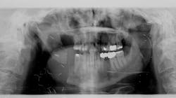
In March 2013, a then-70-year-old male presented to our office for a new-patient exam with a specific request to have an “irritated” tooth in the lower right evaluated. The patient disclosed a previous history of skin cancer in 2007, with a lesion removed from his cheek and subsequent loss of a salivary gland on the right side. A large visible scar over the right masseter area was present, and the patient explained that the surgery had left him with limited ability to open his mouth and care for the teeth on that side. Tooth no. 31 showed significant bone loss on the distal aspect but was otherwise solid (figure 1).
Since the patient went through radiation treatment of the area with his previous surgery, it was decided to attempt localized scaling and root planing, along with frequent recare visits, to help retain no. 31 rather than extract it.
Surgical treatment
In the fall of 2014, our office was contacted by the head/neck cancer team at Virginia Mason Hospital to evaluate the stability of the patient’s tooth no. 31 and determine whether or not it could be removed while they performed a tumor resection of the mandible in that area.
Surgery was performed to remove a squamous cell carcinoma lesion along with tooth no. 31 in November 2014. Initial healing went well for our patient, but as time went on, an ulcerated area distolingual to no. 30 appeared and wouldn't heal. The patient also experienced swelling off and on in the area. By late February 2015, bone had begun to protrude through the tissue, along with severe pain for our patient.
Diagnosis
A diagnosis of osteoradionecrosis of the jaw (ORNJ) was determined. ORNJ describes the process whereby irradiated bone undergoes necrosis and becomes exposed through soft tissue due to therapeutic radiotherapy for head and neck cancer.1 ORNJ most commonly occurs in the mandible at a rate of 5%–20%.1
Multiple surgeries
Once again, our patient returned to Virginia Mason Hospital for surgery. Following his second surgery to remove necrotic bone, the patient continued having issues with limited opening. He sought help from a physical therapist, who offered some improvement using an OraStretch device.
During one of these exercises in mid-April, the patient heard a loud pop and experienced immediate, severe pain. Right away, he went to our local oral and maxillofacial surgeon, who found a horizontal fracture of the ramus just above the angle of the mandible. The previous resected area distal to no. 30 appeared to still be holding, and all teeth fit approximately in occlusion.
By mid-May 2015, things continued to deteriorate for our usually upbeat patient. He was showing signs of exhaustion and losing weight. A new area of ORNJ was growing lingual to no. 30, the patient was in constant pain, and the posterior right side of his face appeared to be sinking in.
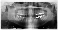
A panoramic radiograph showed the previously resected area was now fully displaced, and the musculature was causing rapid changes in the rotation of the two pieces away from each other (figure 2). The patient now had a fistula draining from the lower border of the mandible through the skin.
Another surgery was planned with the hopes of fixating the mandible back together and removing the area affected by ORNJ and tooth no. 30. The latter two procedures were performed, but the difficult decision of fixation was decided against due to the patient’s poor healing in the past.
After this surgery, our patient felt much relief and had an amazing ability to accommodate using only his left side to chew. The only area of pain came from his now-displaced coronoid process, which was pushing into the approximate area of the maxilla. Unfortunately, the draining fistula would not go away. The patient was treated with several rounds of antibiotics, multiple visits to aBy the spring of 2018, the ORNJ had returned. Another surgery was performed. Another tooth was removed (no. 29), along with more bone.
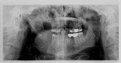
Thankfully, this time surgery was a success. The fistula is gone, and there have been no new ulcerated lesions to date (figure 3).
About the Author
Craig Harder, DDS
Craig Harder, DDS, a native of Spokane, Washington, is a 1995 graduate of the Creighton University School of Dentistry. He practices in Moses Lake, Washington, and previously served as the Grant County Health Department's dental liaison as well as Access to Baby and Child Dentistry champion.


