Seeing is believing: Benefits of using a phase contrast microscope in dental hygiene
Ninety percent of the US population has some level of gum disease, yet most of our patients receive a prophylaxis when they come for their dental hygiene recare appointments. As statistics prove, too often we are performing prophys on patients who have some level of periodontal infection. We scale and polish, stress more flossing and electric toothbrushing, and then recommend that the patient return in six months. Bleeding gums are “the norm.” But prophylaxis by itself will not help a patient heal. Biofilm repopulates within 24 hours,1 as shown by Dr. Bill Costerton,2 the father of biofilm research. Many of our red and orange complex pathogens are resistant to scaling.3
If our patients are doing their best and dental diseases remain, what else can we do?
Seeing is believing
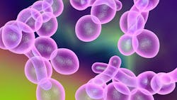
I use a phase contrast microscope in my dental hygiene practice. We show our patients the bacteria, yeast, and parasites that are living within their subgingival plaque biofilm using a microscope projected on a video monitor. A pathological biofilm has a very different appearance compared to a healthy biofilm. Patients of all ages understand that the more frenzied and active the slide appears, the more destructive and aggressive their periodontal disease could become. Parasites, spirochetes, and numerous white blood cells do not belong in a healthy biofilm or a healthy mouth.
Every patient gets a microbiological assessment at every dental hygiene recare appointment. It takes just moments to make the slide. Once the slide is done, I place it to the side of my tray where it needs to rest for at least 10 minutes. I continue with my dental hygiene diagnosis and the rest of the appointment. My patient and I view the slide together near the end of our time. Next, I complete my oral hygiene and self-care recommendations, taking into consideration the results from the bacterial biofilm examination.
New patients exclaim they have never seen their plaque biofilm in action before, and they want their entire family to come in for examinations. Patients discuss their previous efforts to heal and their home self-care tools. But once they see the activity on the slide, they understand even better that string, nylon bristles, and sticks will not reach where these “bugs” live.
Our patients receive individualized instructions to address their dental disease. We often recommend oral irrigation with medication. The goal is to get to the root of our patients’ dental issues, so we include laser therapy, nutrition counseling, airway assessment, ozone therapies, perioscopy, and more in-depth oral self-care instructions in our treatment plans.
Patients return eager to see how effective their home-care efforts are in healing their infections. We can measure progress by looking at the biofilm. The beauty of using a microscope is in seeing the bacteria in real time. Observing the bacteria move, wiggle, and ooze across the screen motivates the patient unlike anything else. We can lecture, demonstrate, beg, and share lab reports, but these have little impact compared to placing a slide on the screen. When patients start asking questions and want to know what they need to do now to make this better, they own their health journey. They are on board and willing to do the work needed to change the pathologic biofilm to one of health.
The best part is when patients return for what we call a tissue and microscope reevaluation follow-up appointment or “tissue check,” and they witness the reduction in bacteria. The reward patients receive by seeing the improvements in both the tissues and then on the video screen make all their hard work worth it. It takes teamwork, and patients have a greater appreciation for the hard work it takes to get there.
Oral-systemic connections
While at the microscope, I share research studies connecting oral pathogens to other body parts, such as the brain,4 heart,5 joints,6 unborn babies,7 and the lungs.8 Patients ask how the pathogens travel to these far-flung body parts. I remind them of the bleeding I just showed them and let them know that it takes the bacteria 60 seconds to reach every nook and cranny of their body.9 What happens in the mouth does not stay in the mouth. I explain that they swallow about 2,000 times a day as well as inhale these pathogens into their lungs.
Then we talk about how important it is for their primary care provider (PCP) to be aware of their gum disease; that the entire body is affected by this inflammation and system-wide infection.10 Including the patient’s PCP in the healing program can help boost the patient’s immune system and address the underlying systemic conditions. We know something is happening deeper within the body that is allowing this dysbiotic biofilm to occur. Bleeding gums, decay, and dysbiotic biofilm are but the “canary in the coal mine” for much bigger systemic issues breaking down elsewhere in the body.
I also use salivary diagnostics to determine the quantities of both good and bad pathogens. These reports are important to share with the patient’s PCP. Knowing these infections are systemic and having the physician’s help in finding the causes are critical pieces of the healing puzzle.
History
Dr. Paul H. Keyes introduced me to the microscope many years ago. His statement, “A dental hygienist without a microscope is like a doctor without a stethoscope,” changed the course of my life. Dr. Keyes was the former head of the National Institute of Dental Research for 25 years. He researched and taught microbiologically monitored and modulated periodontal therapy (MMPT). He stressed identifying the bacteria living in the mouth and shared ways to change the biofilm. He strongly advocated that every hygienist should be practicing with a microscope.
If we can’t see it, we can’t treat it. The simplicity of the scope makes it easy to explain periodontal disease. Without it, I would be the oral janitor at best. At worst, I might be spreading the bacteria and infecting the patient, creating an even worse infection.11
Invest in the best
A chairside phase contrast microscope is an investment in patient health, and dental hygienists are the prevention specialists. Let’s go beyond the traditional scrape and polish to be the true healers we’re meant to be. Having the best tools makes dental hygiene diagnosis quick and easy, while at the same time engaging patients in their oral health. The slide takes moments to make but pays back a hundredfold.
We went into health care to be healers, to help people, and to leave a legacy of true systemic health. More than 90% of the population needs this kind of specialized care. Phase contrast microscopy takes total body health care to another level. Seeing the bacteria, sharing tools and prevention protocols, and helping patients heal make being a biological dental hygienist truly rewarding.
Editor’s note: This article first appeared in Through the Loupes newsletter, a publication of the Endeavor Business Media Dental Group. Read more articles and subscribe to Through the Loupes.
References
- Stoep CV. Biolfilm formation over 8 hour time period. May 1, 2014. YouTube video. https://youtu.be/RmwbqzxQjPs
- Lappin-Scott H, Burton S, Stoodley P. Revealing a world of biofilms—the pioneering research of Bill Costerton. Nat Rev Microbiol. 2014;12(11):781-787. doi:10.1038/nrmicro3343
- Slots J, Ting M. Systemic antibiotics in the treatment of periodontal disease. Periodontol 2000. 2002;28:106-176. doi:10.1034/j.1600-0757.2002.280106.x
- Miklossy J. Alzheimer's disease – a neurospirochetosis. Analysis of the evidence following Koch's and Hill's criteria. J Neuroinflammation. 2011;8(1):90. doi:10.1186/1742-2094-8-90
- Bale BF, Doneen AL, Vigerust DJ. High-risk periodontal pathogens contribute to the pathogenesis of atherosclerosis. Postgrad Med J. 2017;93(1098):215-220. doi:10.1136/postgradmedj-2016-134279
- Maresz K, Hellvard A, Sroka A, et al. Porphyromonas gingivalis facilitates the development and progression of destructive arthritis through Its unique bacterial peptidylarginine deiminase (PAD). PLoS Pathog. 2013;9(9):e1003627. doi:10.1371/journal.ppat.1003627
- Boggess KA, Moss K, Madianos P, Murtha AP, Beck J, Offenbacher S. Fetal immune response to oral pathogens and risk of preterm birth. Am J Obstet Gynecol. 2005;193(3 Pt 2):1121-1126. doi:10.1016/j.ajog.2005.05.050
- Scannapieco FA, Bush RB, Paju S. Associations between periodontal disease and risk for nosocomial bacterial pneumonia and chronic obstructive pulmonary disease. A systematic review. Ann Periodontol. 2003;8(1):54-69. doi:10.1902/annals.2003.8.1.54
- Hirschfeld J, Kawai T. Oral inflammation and bacteremia: implications for chronic and acute systemic diseases involving major organs. Cardiovasc Hematol Disord Drug Targets. 2015;15(1):70-84. doi:10.2174/1871529x15666150108115241
- Martínez-García M, Hernández-Lemus E. Periodontal inflammation and systemic diseases: an overview. Front Physiol. 2021;12:709438. doi:10.3389/fphys.2021.709438
- Allen HB, Morales D, Jones K, Joshi S. Alzheimer’s disease: a novel hypothesis integrating spirochetes, biofilm, and the immune system. J Neuroinfect Dis. 2016;7:1.
About the Author

Barbara K. Tritz, MSB, RDH, HIAOMT
Barbara K. Tritz, MSB, RDH, HIAOMT, has more than 40 years of clinical experience in dental hygiene. She is a biological dental hygienist at Green City Dental in Edmonds, Washington, and has a dental laser certification from the Academy of Laser Dentistry. She is also a practicing orofacial myofunctional therapist and the owner of Washington Oral Wellness in Kirkland, Washington. Barbara was the recipient of the 2019 Hu-Friedy/ADHA Master Clinician Award. Visit her blog at queenofdentalhygiene.net.
Updated April 18, 2023
