What you see may not be what you get
There is a phrase that has been made commonplace in the dental industry by Bill Dickerson, founder and CEO of the Las Vegas Institute - “You don’t know what you don’t know.” This is a powerful statement that certainly applies to all aspects of our lives. It is one that hopefully reminds us of how important it is to be a continual student and to strive to learn more and more as we go through our lives - professionally and personally.
In the last few years, an occlusal philosophy that has been around for many years has been reborn. Neuromuscular occlusion has been a gem hidden from the masses of dentists. This rebirth has ignited a fire in the world of occlusion that has found many practitioners re-examining what they had for so many years taken as gospel. The traditional approach to occlusion that most of us have embraced to some degree has centered around great dentists who have served our profession well. Pankey, Dawson, and many others have given the dental community great principles to follow.
What most dentists will tell you though, if they are being truthful, is that occlusion is and has been a “mystery.” It never really made any sense; the few who did understand it made it sound so difficult and almost secretive that only a select few could be admitted to the club. Most dentists subscribe to the GABS occlusal theory - Grind All Blue Spots. Dentists will leave the patients in the bite they have acquired and fit any new dentistry into that bite.
This often works - my, aren’t humans so wonderfully adaptable? - but what do you do for the person who has problems, has occlusal disease, and/or has TMD symptoms but can’t get any help? There are thousands of people who have occlusal disease - many of whom are in regular care with a dentist - and who have life-altering problems that go undiagnosed.
It seems as though the less you know, the more normal our patients appear. Neuromuscular occlusion offers a different approach to people with occlusal disease and head, face, and neck pain and symptoms. By focusing on the muscles instead of bony relationships, dentists can help patients in ways never before imagined. It may take an initial “big step” out of your comfort zone, but no one who has taken the step, learned the principles, and applied them will argue that traditional occlusal theories work better.
There seems to have been a misconception laid on the neuromuscular occlusal philosophy - all it does is open the vertical dimension of occlusion. I would like to go through a case that I find often in my own practice. This case illustrates perfectly how a neuromuscular approach is not only about vertical - which is actually the least important of the six dimensions of occlusion - but placing the mandible in the most opportune myocentric position.
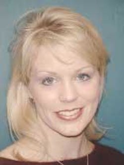
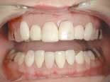
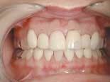
A very pleasant 27-year-old female presented to my office requesting improvements to her failing cosmetic dentistry (figure 1). During her high school years, she had cosmetic dentistry attempted on teeth 6-11 and 22-27. Porcelain veneers were placed without much regard paid to smile design. The veneers were placed with an unknown material but were not bonded to the teeth. This allowed for gross leakage with resulting decay (figure 2).
Along with her cosmetic concerns, this patient reported multiple symptoms she related to a car accident over two years prior. In addition to very tender masseter, temporalis, lateral pterygoid, and sternocleidomastoid muscles, she also reported headaches three or four times per week as well as migraine-type headaches. She described her jaw joint as “sticking” and “locking.” She has been under chiropractic care with limited results. She has also sought the care of several neurologists for help with her migraines and most recently had injections of Botox for her headaches.
In spite of all these attempted remedies, nothing seemed to work for very long. This seemed like an ideal case where dentistry - from a neuromuscular approach - could not only help her with her esthetic concerns, but also help restore her comfort and quality of life. After complete examination and gathering of all pertinent data, a plan was presented and accepted - a full-mouth rehabilitation with a new neuromuscularly derived mandibular position.
The first piece of the puzzle was to find the location of this new position. Using a Myomonitor, a myocentric bite was taken. An upper impression was taken using a Shrinemacher impression tray. This will register the hamular notches, a key in getting the model mounted for proper evaluation. The lower model was mounted against the maxillary model with the myobite and placed on an Accu-Liner for evaluation.
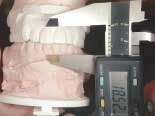
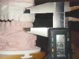
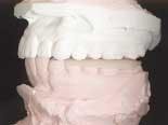
I found something very interesting (figure 3). The anterior vertical dimension was left practically unchanged, a .5 mm difference (figure 4). What did change though was the posterior vertical. Look at how the first molars separated more than 3.5 mm vertically (figure 5). Also changed was the pitch, yaw, roll, anterior/posterior, and lateral position of the mandible.
Opening the vertical anteriorly with this case would have created problems, not fixed them. Also, just think how long the teeth would have become. What an esthetic nightmare! We can see the existing measurement from the CEJ of the maxillary central to the CEJ of the mandibular central was 18.5 mm. With the maxillary centrals measuring 9 mm wide and 11.5 mm long, the existing Shimbashi measurement (CEJ to CEJ) was right where it should be.
This case shows an anterior vertical that is within an appropriate range (19mm+/- 2mm), but a posterior vertical that is lacking. Needing support here to allow the mandible to be in its most optimal position, I made a mandibular orthosis to this exact position determined by the myobite previously taken (figure 6).
This appliance was worn continually for the next four months. During this time, the patient experienced a resolution of all muscle tenderness, headaches, and any symptomatology with which she initially presented. She felt wonderful and was ready to proceed with completing her treatment.
After a smile design had been accomplished, models, pictures, and a prescription for the lab was sent. Returned was a full-mouth wax-up done to the specifics of the bite and esthetics I wanted, as well as stents for the temporaries, a bite stent, and preparation stents, all to help the preparation appointment go as quickly and easily as possible.
The preparation of the full mouth took place in one appointment over about 3.5 hours. This included temporization. Removal of old failing dentistry and the increased interarch space posteriorly allowed for fairly conservative preparations. Several of the teeth still show the remaining central groove (figure 7). The patient left this appointment extremely pleased with her new smile, even though it was only the temporary version. During the time of her temporization, the patient remained symptom-free.
At the seat appointment, all restorations were verified for fit and the shape and shade approved by the patient. Insertion and initial coronoplasty was accomplished in roughly 2.5 hours.
The patient continued to be symptom-free. Several appointments were performed to get exact micro-occlusion to make sure no interferences were present to create any problems.
This process as addressed here in this article seems to be very straight-forward and almost easy, but only after hours and hours of study and preparation can cases like this be accomplished. The marriage of great esthetic dentistry with sound occlusal principles - such as the neuromuscular philosophy - can provide our patients with dentistry and health like never before.
Let’s look now at several before-and-after pictures to show the amazing improvements. At this point, I should take time to thank MicroDental Laboratories for their immaculate work.
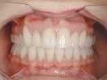
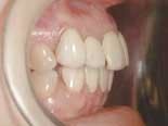
Retracted (figures 8 and 9) - Notice the improvement of the midline, improved shade and esthetics, and the harmonious integration of the Empress restorations with the surrounding periodontal tissues.
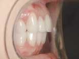
Side retracted (figures 10 and 11) - Notice the improved overjet and overbite as well as the angulation of the maxillary centrals. Also notice the outstanding facial anatomy to create lifelike restorations.
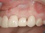
Close-up (figures 12 and 13) - Notice the improved gingival contour, great gingival response secondary to supragingival margins, and, again, great esthetics.
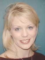
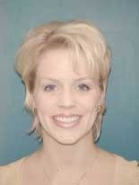
Face (figures 14 and 15) - Now you can see a much more relaxed, happy, and rejuvenated patient with an incredible smile!
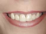
Close up smile (Figures 16 and 17) - Finally, you can see the drastic improvements made in her smile - less gummy, ideal contours, and great color.
Author’s Note: This article was meant to give a cursory overview of how neuromuscular dentistry can be used to achieve full-mouth rehabilitation. Many steps were left out and this type of approach should not be attempted without a thorough understanding of all the principles alluded to here.
Dr. Kevin Winters graduated from the University of Missouri- Kansas City in 1989. After completing a GPR at the University of Louisville-Humana Hospital, he opened a general practice in Claremore, Okla. After developing a successful general practice and being awarded the Young Dentist of the Year award in 1995, Dr. Winters transitioned his general practice to one that concentrates on esthetics and reconstruction. Dr. Winters is one of the original clinical instructors at the Las Vegas Institute. He also lectures and conducts seminars across the nation. Dr. Winters may be reached at (918) 341-4403 or by e-mail at [email protected].
