Endodontic case study: an alternative method for the cleaning and shaping of a mid-mesial canal
Clinician: David Landwehr, DDS, of Madison, Wisc.
Because of anatomical variability, a one-size-fits-all mentality is not possible when cleaning and shaping the root canal system. My instrumentation goal is to enlarge each canal at every level, while respecting the natural canal anatomy and attempting to create cleanliness as well as preserve tooth structure for restoration and function.
For further study |Endodontic case study: Challenges of a bifurcated canal
Chief complaint: Extreme and increasing hot and cold sensitivity from the lower left.
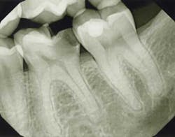
Exam/diagnosis: Tooth No. 19 was hyper-responsive to cold with no linger. The tooth presented with extensive decay into the pulp space, but was asymptomatic to both percussion and palpation. Radiographically, the periodontal ligament space and trabecular pattern were within normal limits.
Pulpal diagnosis: Symptomatic irreversible pulpitis.
Periapical diagnosis: Normal.
PREOPTreatment options: Root canal treatment, no treatment or extraction.
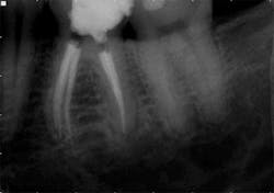
Treatment: Following an inferior alveolar block and intraosseous anesthesia, access was made and caries were removed. After access, the main canals were irrigated with sodium hypochlorite and a glide path was established with hand files to working length. The dentin triangle was removed with Protaper S1 and the final instrumentation was completed using WaveOne. The two canals in the distal root joined to form a broad canal in the facial-lingual dimension and exit a common foramen. After shaping was completed in the mesial root, an isthmus of pulp tissue was identified under the microscope and a mid-mesial canal was identified following exploration of this area. However, the initial orifice was small and the canal path irregular, thereby preventing the establishment of a glide path to the apex. To open the orifice, a series of hand files was used just below the pulpal floor. It was then possible to pass a 10 file to the mid-root and the orifice opening was completed with a Vortex orifice opener size 20/08. The natural glide path was then instrumented to the apex with a 10 file and a Vortex Blue 15/04 was utilized to enhance the shape. A series of Vortex Blue instruments was used to create the final shape of 30/04. In this clinical scenario, Vortex Blue is the ideal rotary instrument system because of the cutting efficiency and combination of cyclic fatigue resistance and flexibility.
The WaveOne shape in the four main canals allowed for obturation with a warm vertical technique, because the heat source could be delivered to within a few millimeters of the apex. However, the constricted shape of the mid-mesial was not amenable to this technique and a GuttaCore carrier was placed to take advantage of the hydraulics and create a dense, three-dimensional seal.
This case illustrates an alternative method of hybridizing the shaping technique based on the natural canal anatomy. In spite of highly variable anatomy in the mesial root, different instrumentation and obturation techniques yielded a final outcome that cannot be distinguished radiographically.
PREOP 1ADDITIONAL ENDODONTIC CASE STUDIES …
Endodontic case study: Hybrid technique on 90-degree curve
Endodontic case study: Healing of a large lesion in a lower molar after proper dental instrumentation and irrigation
Dr. Kinra's case gallery: Soft landing


