Manifestation of a common intraoral autoimmune inflammatory disease

Editor's note: Originally published December 15, 2015. Updated March 1, 2023.
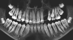
A healthy, 54-year-old, white female presents for evaluation of a painful area on her left and right buccal mucosa.
She reports a whitish-red area bilaterally that is tender to the touch. She states it came up slowly over the last two weeks and nothing seems to make it better. She denies weight loss, fatigue, recent dental work, or trauma in both areas, and there are no extraoral manifestations of these symptoms.
Upon clinical exam, the buccal mucosa bilaterally is erythematous and atrophic, very tender to palpation. There is no cervical lymphadenopathy, and the rest of the oral exam in within normal limits.

Differential diagnosis
- Lichenoid drug reaction
- Oral erosive lichen planus
- Dysplasia
- Oral squamous cell carcinoma
- Mucous membrane pemphigoid
Erosive lichen planus
Oral lichen planus (LP) is a common intraoral autoimmune inflammatory disease. There is also an extraoral cutaneous manifestation of LP that may occur concurrently with the oral form. It may or may not be known to the patient as most of the time the lesion is asymptomatic.
More pathology cases ...
The pathology with multiple manifestations
A patient with a leukoplakic lesion and a noncontributory health history
LP is divided into two categories: the reticular form and the erosive form.1 The reticular form is more common than the erosive form. Typically, the reticular form is asymptomatic to the patient. Clinically, it is characterized by a white, lacy latticework of mucosa found on the buccal mucosa, attached mucosa, and tongue. These white striae of mucosa are referred to as Wickham striae. The erosive form, as outlined in the above vignette, is the more symptomatic form of LP. It is typically characterized by painful, atrophic, ulcerated areas of the oral mucosa.2
Diagnosis of this condition is dependent on the form of LP the patient presents with. The reticular form is usually made on the basis of a clinical presentation. An incisional biopsy can also be performed, or treating the area empirically with a topical corticosteroid and ensuring resolution of the lesion after two to three weeks can aid the clinician in being sure of his or her diagnosis. When the area in question is suspected to be erosive lichen planus (ELP), a biopsy of the lesion is typically advised, not only to ensure the area is in fact ELP, but to rule out dysplasia or a malignancy.
Recommended treatment
Once a diagnosis is made, treatment of the lesion depends once again on the form of LP. Most reticular forms of LP require no treatment, as they are usually asymptomatic and nonprogressive. Routine dental hygiene follow-up is typically adequate to ensure the area is not changing or becoming symptomatic to the patient. ELP typically requires treatment with either a systemic or topical corticosteroid, or a combination of both. New cases with moderate to severe symptoms typically require a short course of systemic prednisone to get the disease under control, followed with maintenance using a topical corticosteroid such as fluocinonide gel (Lidex)2 or triamcinolone in Orabase (Kenalog). Mild cases typically can be managed from the beginning using topical corticosteroids. Follow-up of ELP cases is recommended to ensure the area stays under control with the topical agents, as well as to ensure that there is no malignant transformation of the area or areas of moderate to severe dysplasia that were missed on the biopsy sample that can progress to a malignancy.1,2
References
1.Damm N, Bouquot A. Oral and Maxillofacial Pathology. 2nd ed. WB Saunders; 2002:680-685.
2. Marx RE, Stern D. Oral and Maxillofacial Pathology. Quintessence Books; 2003:156-162.
About the Author
Kevin J. Connor, DDS, MD
Kevin J. Connor, DDS, MD, has a BS in biomedical engineering and earned his DDS degree from Marquette University School of Dentistry. He completed medical school at the Louisiana State University Health Sciences Center, followed by a one-year general surgery internship. He did his oral surgery training in 2010 at LSU and is currently in private practice outside Milwaukee, Wisconsin. Dr. Connor’s surgical interests include dentoalveolar trauma, reconstructive surgery, and surgical treatment of temporomandibular joint diseases.