How to obturate like an endodontist
Would you like your root canal procedure outcomes to be more consistent and successful? Endodontist Dr. L. Stephen Buchanan simplifies the technicalities of obturation using 3-D condensation with the Continuous Wave of Condensation Technique, which he himself developed, along with continuous wave electronic devices and pluggers.
Editor's note: This article first appeared in DE's Breakthrough Clinical with Stacey Simmons, DDS. Find out more about the clinical specialties newsletter created just for dentists, and subscribe here.
THE MOST WIDELY ACCEPTED ROOT CANAL OBTURATION TECHNIQUE THAT ENDODONTISTS USE TODAY is 3-D condensation with the Continuous Wave (CW) of Condensation Technique.
Why we obturate
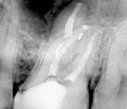
Why does 3-D obturation of root canal systems matter? The biologic rationale for 3-D obturation (root canal systems filled to their full apical and lateral extents) is that we never know for certain whether we have killed every bacterium in any given root canal system. If I have failed to achieve sterility in a root canal system—which is always a possibility despite my best efforts—I can still achieve successful healing if I entomb any remaining infectious agents with sealer and gutta-percha (figure 1). Herbert Schilder, DDS, was the first to describe a predictable obturation method—his Vertical Condensation Technique, also described as the vertical compaction of warm gutta-percha (1)—that could fill any lateral canal complexity that was cleaned beforehand.
Figure 1: Maxillary molar with an MB2 canal that bifurcated, mid-root, from the MB1 canal, made a 90-degree turn, and bifurcated again in its apical 3 mm. A canal form such as this is unlikely to be known before or during root canal treatment, presenting more than 7 mm of canal space that was unnegotiable, unshaped, and most certainly not sterilized during irrigation procedures. This is a case that succeeded only because the inevitable bacterial remnants were entombed by filling material during 3-D obturation.
Continuous Wave of Condensation Technique
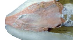
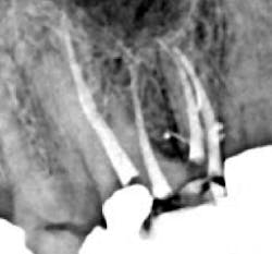
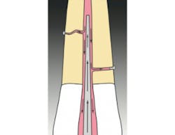
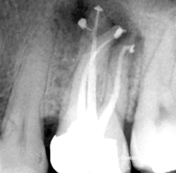
In 1989, I conceived of a way to radically simplify 3-D obturation.This new method collapsed Schilder's Vertical Condensation Technique of three-to-five heating and packing steps requiring five minutes per canal, into a downpack that required just two procedural steps and less than 15 seconds per canal to complete. While it was my intention was to simplify Schilder's procedure, the surprise result was a "centered" condensation technique that, despite the huge reduction in time and skills needed, actually provided superior obturation results that moved more gutta-percha into lateral complexities than vertical condensation (figures 2, 3, 4a, and 4b). (2)
Figure 2: My first Continuous Wave of Obturation result in a maxillary molar. Note the significant mid-root lateral canal filled off the MB2 canal, the mid-root isthmus filled between mesiobuccal canals, and the lateral canal filled off the MB1 canal.
Figure 3: Illustration of "centered" condensation, with its streaming effect. The best analogy for the CW Technique is that it is the inverse of analog impressioning. The impression tray becomes the electric heat plugger, the heavy-bodied impression material is like thermo-plasticized gutta-percha, and the thin-bodied material is analogous to sealer.
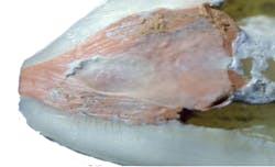
Figure 4a: Sagittal dissection of the mesial root of an extracted mandibular molar, showing 3-D obturation result with the Continuous Wave of Obturation Technique. Note the gutta-percha and sealer completely filling the isthmus between the MB and ML canals. (Courtesy of Robert Sharp, DDS)
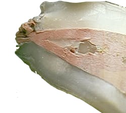
Figure 4b: Sagittal dissection of MB root of an extracted maxillary molar, showing a similar 3-D obturation result with the Continuous Wave of Obturation Technique. Note the gutta-percha and sealer completely filling the isthmus between the MB1 and MB2 canals. (Courtesy of Gary Carr, DDS)
Continuous wave electronic devices
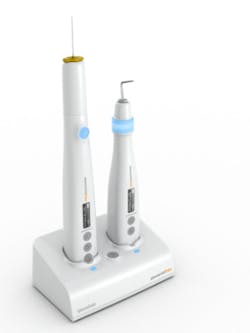
Figure 5: The elements free Cordless Continuous Wave Obturation devices on their charging stand
Over the years, many devices have been created to assist in the delivery of warm vertical compaction techniques: Touch 'n Heat, System B, Elements Obturation Unit (EOU), to name just a few. However, the elements free (EF) devices (Kerr Dental) are the first cordless units I have used that work exactly like the corded EOU unit operates, which means the electric heat pluggers reach full temperature within 0.5 seconds, and the gutta-percha backfilling devices work effortlessly without ratcheting problems (figure 5).
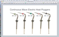
Figure 6: The full array of Continuous Wave of Obturation Electric Heat Pluggers
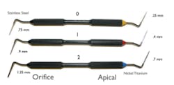
Figure 7: Buchanan CW Hand Pluggers 0, 1, and 2 with color ring indicators at the nickel titanium apical plugger ends. Note the NT plugger diameters of .25 mm, .4 mm, and .7 mm, respectively, and the stainless-steel orifice plugger diameters of .75 mm, .9 mm, and 1.35 mm, respectively.
Continuous wave pluggers
The CW Technique is used with electric heat pluggers (EHP) and hand pluggers (figure 6). EHPs are available in five different sizes: .04, .06, .08, .10, and .12 tapers. All but the .04 EHP have tip diameters of 0.5 mm; the .04 is intended to downpack through minimally invasive endodontic (MIE) shapes, so it has a smaller tip diameter of .3 mm. The objective is to use the largest EHP that fits within the desired 4 mm to 6 mm (from the terminus) depth, because it will bind more closely at the orifice level of the canal, creating a maximal wave of condensation pressure during the downpack.
The three hand pluggers (figure 7) are sizes 0, 1, and 2, with have yellow, red, and blue colors, respectively, at the apical plugger end. Size 0 is used when the 30-.04 EHP is selected for use in minimally shaped canals or canals with severe curvatures in their coronal halves. The size 1 plugger is used for downpacking most small canals that are typically prepared to size 20-.06, 30-.06, and 40-.06 shapes (fit with .06 or .08 EHPs). The size 2 plugger is used for medium and large canals shaped to 30-.08 or larger. Its NT apical end is .7 mm in diameter, and its SS orifice end is 1.35 mm in diameter.
Conclusion
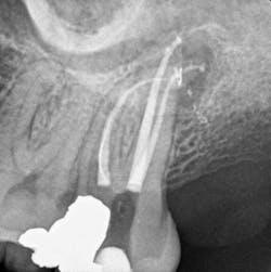
GT hand and rotary files took six years to develop. Conversely, within a month of conception, I had the first prototype electric heat plugger, and it worked well the first time I used it to do a Continuous Wave downpack. Ironically, in product development, it isn't always the failures that we must deconstruct and design around. With the CW Technique, it took more than two years to deconstruct that success to fully understand the power of "centered" condensation. Since then, I have been gratified to see the widespread adoption of this technique, making Schilder's filling results accessible to endodontists and general practitioners alike (figures 8a and 8b). To all who have adopted this technique, you have my deepest gratitude. But please, just a humble request, don't call it Vertical Condensation.
Figure 8a: Maxillary molar obturated with the Continuous Wave Technique. Note the significant lateral canal off the palatal canal. (Courtesy of Dr. Giuseppe Cantatore; Rome)
Figure 8b: Maxillary molar obturated with the Continuous Wave Technique. Note the multiple lateral canals and the long MB2 fin off the MB1 canal. (Courtesy of Constantinos Laghios, DDS, MS; Athens)
Author's note: elements free is a trademark of Kerr Corporation. All other trademarks are the property of their respective owners.
References
1. Schilder H. Filling root canals in three dimensions. Den Clin North Am. 1967;723-744.
2. DuLac KA, Nielsen CJ, Tomazic TJ, Ferrillo PJ Jr, Hatton JF. Comparison of the obturation of lateral canals by six techniques. J Endod. 1999;25(5):376-380.
Editor's note: This article first appeared in DE's Breakthrough Clinical with Stacey Simmons, DDS. Find out more about the clinical specialties newsletter created just for dentists, and subscribe here.
