Summary of furcation defects for dental hygiene exams
Interview with Claire Jeong, RDH, about her website, StudentRDH.com.
By Claire Jeong, BS, MS, RDH, and Leon Yu, DMD
As a #RDH or #futureRDH, we need to know about the periodontium, the base structure that supports the teeth. Furcation assessments are one of the necessary measurements we take to evaluate the patient's periodontium because knowing how much bone the patient has lost helps us understand the oral health of the patient. If furcation is present, the dental hygienist should clean the area.
Let's go over furcation classifications as part of the mini-review series for the dental hygiene board exams. If you've already passed, this review will be just for fun!
Furcation Definition And Detection1
Furcation is the anatomical area where the roots divide. Therefore, furcation defect (also called furcation involvement) refers to bone loss at the branching point of the roots. Furcation can only be present on multi-rooted teeth, not single-rooted teeth.
Diagnosis of the furcation involvement is based upon probing and visual detection. Although a straight probe may be used, detection of subgingival furcation is facilitated by the use of the curved Nabers probe. Radiographic images can also help detect furcation (e.g., a radiolucency between the roots indicates a furcation defect of Class II and above) but are not completely reliable as overlapping of tooth structures can mask the bone loss.
Glickman’s Classification2
Glickman’s classification of furcation involvement are as follows:
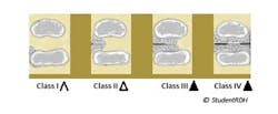
- Class I: Curvature of the concavity between the roots can be detected with the probe tip but it cannot enter the space.
- Class II: Probe penetrates into the furcation, but does not completely pass through to the other side.
- Class III: Probe passes completely through the furcation but is not clinically visible because the soft issue still fills the furcation defect.
- Class IV: Probe passes completely through the furcation and the entrance to the furcation is clinically visible because of gingival recession.
If you are taking the dental hygiene national or regional exams, note that the severities of furcation defects are indicated with triangle signs as shown in the image above.
Furcation Detection According To The Number And Location Of Roots
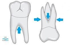
Let’s add one more lesson to this review. Furcation defects should be inspected from different angles according to the number of roots the tooth has.
- Mandibular teeth: bifurcated (mesial and distal roots). Potential furcation defect on the mid-facial and mid-lingual aspects of the tooth.
- Maxillary teeth: trifurcated (mesiobuccal, distobuccal, palatal roots). Potential furcation defect on the mid-buccal, mesial, and distal aspects of the tooth.
- Maxillary first premolar: can be bifurcated (buccal and palatal roots). Potential furcation defect on the mesial and distal aspects of the tooth.
Review the example below.
Q: If the Naber’s probe passes through the furcation to the other side but the furcation is not clinically visible, which type of furcation does this describe?
A. Class I
B. Class II
C. Class III
D. Class IV
Answer: C. Class III. The key words are “to the other side” and “not clinically visible.” From those clues, we can conclude that the tooth has Class III furcation. If the furcation was clinically visible, Class IV would be the answer.
It is extremely important to remember the furcation classifications by heart. The dental hygiene board examination may challenge you to apply your knowledge to detect furcations from radiographs and dental charts to solve case studies. So take the time today to memorize the basics about furcation from this article, and become more ready for your big day!
Claire Jeong, MS, BS, RDH, is the founder of StudentRDH, a review solution for the National Dental Hygiene Board Exams and Local Anesthesia Dental Hygiene Board Exams. She graduated from MCPHS University, Forsyth School of Dental Hygiene; and is a member of Sigma Phi Alpha, the dental hygiene honor society. Claire has a passion for creating educational solutions for the dental hygiene community and is currently helping students from all around the United States and Canada pass their dental hygiene board examinations. Claire is licensed in the United States and Canada. She provides personalized mentorship at StudentRDH and can be reached at [email protected].
Leon Yu, DMD, is the clinical instructor in periodontics at the University of Alberta School of Dental Medicine. Dr. Yu received his doctor’s and Post-doctorate degree in Periodontics from Boston University Goldman School of Dental Medicine. Dr. Yu is a diplomate periodontist certified by the American Board of Periodontology. He is currently in private practice specializing and limited to periodontics and implants. He is also clinical instructor in Periodontics at the University of Alberta.
References
- Nield-Gehrig JS, Willmann DE. Assessment for Clinical Decision Making. In: Foundations of Periodontics for the Dental Hygienist. 2nd ed. Baltimore, MD: Lippincott Williams & Wilkins; 2008:188.
- Newman MG, Takei H, Klokkevold PR et al. Furcation: Involvement and Treatment. In: Carranza’s Clinical Periodontology. 12th ed. St. Louis. MO: Elsevier Saunders; 2012:623.


