Ultra Cone Beam CT Imaging

Changing implant dentistry
By Ronald A. Bulard, DDS, Lacey Clampet, & Horst F. Bruning, PhD
More and more dentists are using X-ray Cone Beam CT (CBCT) Scanners for patient imaging and diagnostics as a new and vital part of their practices. The result is advanced, state-of-the-art volumetric images that increase the quality and accuracy of radiographic dental care. When using CBCT imaging, clinicians will have the most accurate anatomic information to plan the placement of dental implants in optimal sites by using technologically advanced digital imaging devices.
Computerized tomography is advancing rapidly. The imaging source-detector and the method of data acquisition distinguish cone beam tomography from traditional CT imaging. Traditional CT uses a high-output rotating anode X-ray tube, while cone beam tomography utilizes a low-power, medical fluoroscopy tube that provides continuous imaging throughout the scan. Traditional computerized tomography records data with a fan-shaped X-ray beam onto image detectors arranged in an arc around the patient, producing a single slice image per scan. Each slice must overlap slightly in order to properly reconstruct the images. The advanced cone beam technology uses a cone-shaped X-ray beam that transmits onto a solid-state area sensor for image capture, producing the complete volume image in a single rotation. The sensor contains an image intensifier and a CCD camera, or an amorphous silicon flat panel detector.
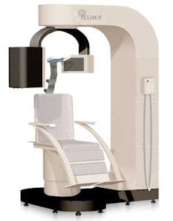
The single-turn motion image-capture used in cone beam tomography is quicker than traditional spiral motion, and can be accomplished at a lower radiation dose as a result of no overlap of slices. This type of imaging exposes a patient to less radiation than traditional CT scanners. Manufacturers are designing Cone Beam Scanners with the physical space available in clinics and the patient’s comfort in mind. For example, upright seating is used in CBCT scanners with the X-ray tube and panel detector rotating around the patient’s head.
Advantages of using CBCT imaging in implant dentistry
Cone Beam CT imaging software helps clinicians visualize a patient’s anatomy in a clinically meaningful way. CBCT imaging is the ideal radiological modality for implantology due to the high quality of the produced images, software capabilities, ability to manipulate and use information for surgical planning, and lower doses of radiation exposure. In addition, the surgery may be performed on the computer first and the results put on a 3D printer to produce a solid model for fabrication of surgical guides and stents.
CBCT imaging allows clinicians to illustrate recommended implant treatment plans to patients. The technology of the cone beam CT scanner impresses patients and aids in the selling of optional, out-of-pocket implant treatment plans.
A CBCT scanner simplifies treatment planning and helps achieve implant acceptance. A CT scan also enables dentists to evaluate the quality and quantity of available bone. Possessing an in-office cone beam scanner eliminates scheduling a patient scan at a hospital or other imaging center. Immediate images make it more likely that patients will accept implant treatment plans.
Cone beam volumetric tomography (CBVT) imaging provides a necessary perspective in many cases that require a lot of information. Examples of applications of CBVT in diagnosis and treatment planning are TMJ conditions and disorders, jaw pathology, orthodontic evaluation, airway analysis, impacted teeth, periodontal defects, and endodontic problems.
The next generation of CBCT scanners
Cone beam volumetric imaging is a recent and fast-growing addition to the dental industry. In 2004, the number of options for cone beam scanners more than quadrupled, and the future of CBCT imaging looks even brighter. The next generation of CT imaging in dentistry is imminent. We, the authors, predict that within the next six months, a newly developed CT scanner with enhanced features will be available to the dental world. The enhanced CT scanner has evolved into a new classification; these new machines will be classified as “Ultra Cone Beam CT Scanners.”
Ultra CBCT imaging provides important information about the three-dimensional structure of blood vessels, nerves, soft tissue, and bone. Imaging helps clinicians plan treatment strategies with the greatest chance for success. A great advantage of using ultra cone beam images when diagnosing patients and evaluating treatment options is the clinician’s ability to visualize the patient’s anatomy in 3-D, instead of flat, two-dimensional images.

Three-dimensional software simplifies the assessment of anatomic considerations and prosthetic goals for implant planning. Three-dimensional software accurately determines bone dimensions and identifies bone quality through density shading. This advanced software determines information about the axis orientation of the alveolar bone for successful implant alignment. It identifies common internal anatomy needed to evaluate implant placement including the jaw boundaries, adjacent teeth, nasal fossa, mandibular canal, maxillary sinus, mental foramen, and incisive canal. It also detects pathology to be avoided for implant health.
Three-dimensional visualization software can shade images to differentiate varying densities of facial structures. Grayscale shading provides the ability to view the relationships of common internal anatomy. Typical cone CT imaging renders an 8-bit grayscale (256 shades) or 12-bit grayscale (4,096 shades). IMTEC Imaging’s new ILUMA Ultra Cone Beam CT Scanner renders images in 14-bit grayscale, providing 16,384 shades. Some versions of volumetric software, like that used in ILUMA, permit the clinician to color code the image by density, further distinguishing anatomical structures. Color-coding the anatomical features enables the clinician to view them while planning implant cases, such as nerves and nasal cavities, and their relationship to the density and length of the mandible or maxilla.
Image data may be obtained for a complete dental/maxillofacial volume or a limited region of interest. With the current generation of cone beam scanners, scan times for these types of images vary from about 40 to 60 seconds for the complete volume and as few as 10 seconds for the regional scan. The next generation ultra cone beam scanners, like IMTEC Imaging’s ILUMA, can take full volume scans in 40 seconds or less.
Amorphous silicon flat-panel detectors, found in some next-generation CBCT scanners, provide orthographic images. These panels reduce optical distortion that is inherent to CCD camera detectors. The images from flat panels are much sharper and dimensionally accurate for correct measurements. The accuracy of images is vitally important to treatment planning, especially in implant surgery cases. Dimensions of sensors (pixels) vary by machine, and some offer two sizes of displays.
Volumetric software programs also enable the clinician to segment the image to evaluate specific anatomical structures of interest. Segmentation literally cuts the volume rendering, conceding top views, side views, and CT slices that produce unlimited axial, coronal, and sagittal views. Ultra Cone Beam CT slices are as thin as 0.1 mm, compared to 1 mm for a conventional fan CT scan. Some software programs on the market, including the one found in ILUMA, allow dentists to transform the digital images into STL format, which is used in the creation of models. ILUMA V-Implant software provides features for the exportation of STL files to 3-D printers, which allows modeling and fabrication of surgical guide stents. Created stents may be strategically placed in images to show the exact angulations and placement of the proposed implant. It is possible to place implants and final restorations the same day using the treatment planning options of the ILUMA V-Works and V-Implant software export features.
Ultra CBCT devices may be used for traditional forms of radiography, in addition to advanced 3-D volumetric renderings. Conventional cephalometric measurements may be obtained through 3-D volumetric images by rendering the image as a 2-D projection resembling a radiograph or a panoramic image. It is also possible to digitize cephalometric points in 3-D, resulting in the introduction of multiple analyses. Ultra cone beam tomography can be easily used in preventative and restorative care cases.
Raising the standard of care in implant dentistry
Ultra Cone Beam CT imaging is quickly becoming the standard of care in implantology; however, the technology is evolving. The software and hardware in CT technology is continually improving, making the cost of an ultra cone beam scanner a deterrent in many private practices.
The current generation of cone beam scanners ranges from $170,000 to $300,000 each with maintenance each year as much as $30,000. As a result, they’re generally available only to imaging centers, group practices, hospitals or large clinics. The high cost effectively reduces patient and clinician access to this advanced technology. Also, the cost of an outside imaging examination is usually greater than that of inside imaging examinations. We believe that if clinicians could obtain access to CBCT technology without committing a great amount of business capital into a depreciating technology, the use of cone beam scanners would rise exponentially.

As with most developing technology, prices of Ultra CBCT scanners will invariably decrease as the computer-aided learning evolves. Uniquely, IMTEC Imaging developed a marketing plan that results in a profit from the very first scan a clinician orders, and eliminates the hazards of technology depreciation. Instead of selling the ILUMA to clinicians, the company will simply place scanners in the clinics of clinicians. The patron pays a minimal refundable installation and training deposit, then pays the vendor each time the machine is used. This results in a significant savings compared to purchasing a machine or outsourcing the imaging work.
Ultra cone beam tomography is quickly increasing the quality and accuracy of radiographic dental care. Used in a wide variety of cases requiring additional information for treatment planning and detection of possible anatomical problems, Ultra CBCT scanners are rapidly changing the field of dentistry.
Utilizing Ultra CBCT technology enhances patient trust and increases treatment acceptance. Possessing an Ultra Cone Beam scanner decreases implant case completion time while increasing the chances of implant success. Within the next six months, ultra cone beam tomography will be accessible and profitable for private practices. As a result, Ultra CBCT imaging is rapidly becoming a standard of radiographic care in dentistry.
Dr. Ronald A. Bulard graduated from the University of Oklahoma School of Dentistry. Lacey Clampet is in public relations at IMTEC Imaging, LLC, Ardmore, Okla. Horst F. Bruning earned a PhD in particle physics at CERN, Geneva, Switzerland. He resides in Pleasanton, Calif.