A laser technique for frenum removal
By Eric Bornstein, DMD
Labial frenal attachments are thin folds of mucous membrane with enclos-ed muscle fibers that attach at the lips to the alveolar mucosa and underlying periosteum. A frenum can become a significant problem if tension from lip movement pulls the gingival margin away from the tooth, or if the tissue inhibits the closure of a diastema during orthodontic treatment.
Because many dental patients are increasing their roles in the periodontal therapy decision-making process, and would rather not suffer the pain and discomfort generally associated with mechanical periodontal surgery, an alternative approach to traditional periodontal surgical procedures is warranted. By combining two different lasers (Er:YAG and SuperPulsed CO2), the general dentist can perform a frenectomy procedure to aid orthodontic treatment simply and predictably with controlled delivery of laser energy making use of the unique characteristics of each wavelength. This can be accomplished by a general dentist trained in laser use and photobiology using the OpusDuo (OpusDent USA), without the need for a periodontal specialist and with minimal trauma to the surrounding tissues, minimal pain, and expedited healing.
Traditional frenectomy surgery
Traditional frenectomy surgery can be accomplished in one of three different ways.
- Simple excision using a narrow elliptical pattern around the frenum down to the periosteum, with a dissection of the muscle from the periosteum and a reapproximation of the wound.
- The Z-plasty technique, which is an extension of the simple excision where the two pointed flaps are rotated and approximated to close the wound.
- A localized vestibuloplasty, with secondary epithelialization of the wound.
The above three approaches all have the following in common — the need for incisions with steel scalpels, mechanical excision of the muscle attachment from the periosteum, placement of sutures, significant postoperative discomfort, and delayed healing. When the benefits and risks of alternative treatment modalities are taken into account for this mechanical surgical procedure, minimally invasive laser-assisted soft-tissue excision and ablation is a very attractive technique to accomplish a simple frenectomy procedure.
Soft-tissue laser procedures
Previously, the Continuous Wave or Pulsed CO2 laser has been used as a surgical tool for precise cutting and coagulating of oral soft tissues. Surgeons using older Continuous Wave or Pulsed CO2 lasers for soft-tissue excisional procedures have previously needed to work very quickly to minimize unwanted thermal damage to the surrounding tissues and bone. If the soft-tissue procedure is accomplished with a dual approach (Er:YAG and SuperPulsed CO2), the potential for thermal damage is greatly minimized, and the window of available working time is greatly expanded. This can be accomplished because of the unique characteristics and photobiology of the Er:YAG laser tissue interaction. With the addition of the Er:YAG laser, the general dentist is presented with a less stressful and more beneficial treatment protocol for soft-tissue excisional procedures than previously available with the older continuous wave or pulsed CO2 lasers alone.
Er:YAG and CO2 thermodynamics and photobiology
When dealing with lasers that are highly specific for water absorption, there are two basic concepts to keep in mind — thermal expansion and heat transfer through conduction. Water is the dominant target chromophore (absorptive tissue element) for both CO2 and Er:YAG laser energy. The photobiology of each wavelength is unique and has distinct benefits for the practitioner. If these characteristics are clearly understood and used to maximize desired effects while minimizing collateral tissue damage, elegant and simple surgical procedures can be accomplished.
The Er:YAG laser emits infrared optical energy at 2.94 microns in the mid-infrared electromagnetic spectrum. Er:YAG laser energy currently has the highest absorption peak in water of any commercially available, FDA-approved dental laser. When operating in a pulsed mode with 200-400ms pulse widths, 5-40 microns (1 micron = 1,000 millimeters) of thermal mechanical tissue ablation (tissue removal) with as little as 5 microns of residual thermal damage is the norm. These unique characteristics of the Er:YAG laser allow for very narrow layers of thermal mechanical tissue ablation with minimal collateral thermal consequences.
The CO2 laser emits infrared optical energy at 10.6 microns in the far infrared electromagnetic spectrum. When operating in a Superpulsed mode with a CO2 laser, many of the problems associated with unwanted thermal residual damage of older Continuous Wave or Pulsed CO2 lasers can be avoided. In the Superpulsed mode, tissue penetration depths of 60-75 microns of thermal tissue vaporization can be expected with thermal hemostasis and coagulation occurring to a depth of 75-150 microns.
Once the unique characteristics of each laser and the tissue interactions associated with them are clearly understood, the benefits of a dual approach to the frenectomy procedure become clear. Because the penetration depth of superpulsed CO2 lasers can approach three times that of Er:YAG lasers, and thermal interactions can approach a depth of 15 times that of Er:YAG lasers, the Er:YAG can and should be used for the excisional mucogingival surgery in this procedure. As the Er:YAG laser only interacts thermally within 5 microns of the tissue target, while the superpulsed CO2 has a much greater optical and thermal penetration depth, the superpulsed CO2 can and should be used for hemostatic purposes in this procedure.
Dual approach case study
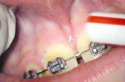
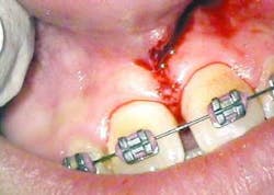
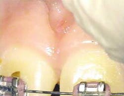
The following is a case report of one such procedure using the Opus Duo, a dual Er:YAG and CO2 dental laser system. This case begins with the diagnosis of a frenal attachment connected to the interdental papilla between maxillary central incisors (No. 8 and No. 9). The muscle attachment is bulky, and would inhibit further orthodontic correction. This patient was referred by her orthodontist for a minimally invasive laser frenectomy procedure before the next stage in her orthodontic treatment (figure 1).

null
null
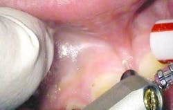
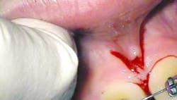
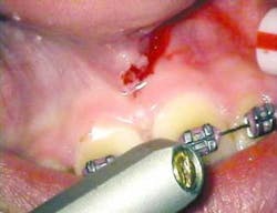
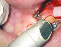
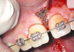
After infiltration of 1cc of local anesthetic solution, the Er:YAG laser (OpusDuo, OpusDent USA) was used at 350mj with a 1,000-micron, contact sapphire tip and heavy water spray to gently ablate the frenal attachment and underlying muscle tissue down to the level of the periosteum (figures 2, 3, and 4). Once this is accomplished, there is a small defect in the vestibular mucosa and interdental papilla where the frenal attachment and muscle once was. There is no charring, no burned tissue, and free bleeding in the site (figure 5).
null
null
Venugopalan et al. described the Erbium tissue ablation mechanism in very elegant terms in 1995. He wrote, "If water is the dominant chromophore, the mechanical integrity of the extracellular matrix (collagen) is not targeted directly. To achieve material removal, the heated water (intracellular steam) must expand first straining and then fracturing the extracellular matrix components (collagen)." Hence, because Er:YAG energy has such a high absorption peak in water, the thermal damage to the tissue is kept to a bare minimum as the thermal mechanical ablation takes place (i.e. no char). The healing capacity of the laser-irradiated tissue when looked at with this logic is profound. Neev et al., in a thermo-optical skin conditioning study, stated that less thermally induced damage means less collagen remodeling is necessary. With less collagen damage and remodeling, faster and easier healing with minimal scarring is the norm.
null
null
Once the Erbium part of the procedure is completed, the Superpulsed CO2 (OpusDuo, Opusdent USA) is then used to simply coagulate and gently cauterize the bleeding site. With this approach, the practitioner can immediately pick up a focused 90-degree handpiece and apply 1.5 watts in the Superpulsed noncontact mode. This is accomplished with a circular sweeping pattern to gently coagulate the superficial bleeding layer to a depth of 75-100 microns (figures 6 and 7).
null
null
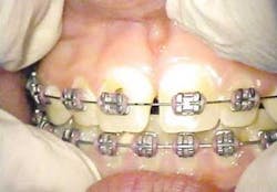
With the dual approach to this procedure, healing at 16 days is profound and complete (figures 8 and 9). Any excess thermal deposition or damage to the tissue is avoided when both lasers are used, and the problematic frenum attachment inhibiting further orthodontic correction is completely removed.
null
Conclusion
As laser use in the practice of general dentistry continues to increase, there will be many procedures that were once done by traditional mechanical methods, but will now be seen through laser safety glasses. If a clinician decides to use a laser for a dental procedure, he or she needs to fully understand the character of the wavelength being used, and the thermal implications and limitations of the optical energy. The above surgical case combined the best characteristics of two different dental wavelengths to create an easier procedure that a general dentist could certainly accomplish. This minimally invasive laser-assisted frenectomy was accomplished with minimal anesthesia, minimal discomfort, no sutures, no antibiotics, no postop visit, and great patient satisfaction.
References
- Newman M, Takei H, Carranza F: Carranza's Clinical Periodontology, ed 9. Philadelphia, WB Saunders Co, pp 870-872, 2002.
- Matthews D, Smith C, Hanscom S, Tooth Loss in Periodontal Patients. Journal of the Canadian Dental Association. Vol 67: 207-210, 2001.
- Peterson L, Ellis E, et al: Contemporary Oral and Maxillofacial Surgery, ed 3. St. Louis, Mosby, pp 312-317, 1998.
- Clayman L, Kuo P: Lasers in Maxillofacial Surgery and Dentistry, New York, Thieme, pp 63-65, 1997.
- Lanigan, S: Lasers in Dermatology, London, Springer, pp 57-79, 2002.
- Venugopalan V, et al: Pulsed Laser Ablation of Tissue: Surface Vaporization or Thermal Explosion?, Wellman Laboratories of Photomedicine, Harvard Medical School, SPIE Vol. 2391, 1995.
- Neev J, et al: Thermo-optical Skin Conditioning: A new method for thermally modifying skin conditions. Lasers in Surgery: Advanced Characterization, Therapeutics, and Systems XII, SPIE Vol 4609, 2002.
Dr. Eric S. Bornstein graduated from the University of Vermont with a bachelor's degree in 1988, Tufts University School of Dental Medicine in 1992, and the Maimonides Medical Center General Practice Residency program in Brooklyn, N.Y., in 1993. He practices general, implant, and laser dentistry in Natick, Mass., along with operating the Metrowest Maxillofacial Imaging Center at the same location. He can be reached by e-mail at [email protected].