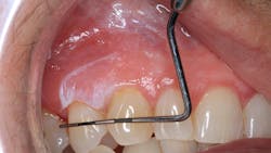
Pathology case: Lesion's dysplastic nature gives rise to concern
Editor's note: Originally published in August 2017. Updated December 1, 2024.
Case presentation
A healthy 66-year-old female presented for a recare exam. The patient's health history was noncontributory except for a hip replacement in the last year.
Clinical exam
Clinical examination revealed a white, corrugated, irregular-bordered lesion on the attached tissue on the upper right side between teeth nos. 5 and 6. The lesion measured approximately 9x6 mm. The lesion was not tender to touch or palpation, and it was not able to be removed or scraped off.
The patient was unaware of the presence of the lesion.
The patient presented two weeks later after the initial discovery of the upper right vestibular lesion. For the most part, the lesion had not changed, except that it had extended back toward the first molar to the mucolabial fold area. The patient was referred to an oral surgeon for assessment and biopsy.
Impressions of the lesion
- No real clear etiology. The patient presented with a clean health history.
- The dysplastic nature of the lesion, with it presenting as a large leukoplakic patch over erythematous tissue, does give rise to concern and is overall more worrisome. Lack of pain and tenderness does not negate its potential severity.
Differentials
- Lichen planus
- Carcinoma (in situ)
- Benign hyperkeratosis
A biopsy was scheduled for a definitive diagnosis.
Regrettably, due to scheduling conflicts and the timing of this writing, the patient has not yet had a biopsy.
Reference guide for leukoplakic lesions
A short background on leukoplakic lesions can be found in a previous pathology case as a reference and general guide to follow.
About the Author
Stacey L. Gividen, DDS
Stacey L. Gividen, DDS, a graduate of Marquette University School of Dentistry, is in private practice in Montana. She is a guest lecturer at the University of Montana in the Anatomy and Physiology Department. Dr. Gividen has contributed to DentistryIQ, Perio-Implant Advisory, and Dental Economics. You may contact her at [email protected].

