Editor's note: Originally published August 16, 2019. Formatting updated November 15, 2022
Case history and presentation
A 66-year-old male presents for his routine exam and periodontal maintenance visit. His health history includes medications for blood pressure and cholesterol.
Clinical assessment
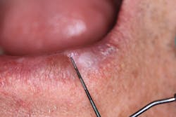
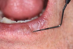
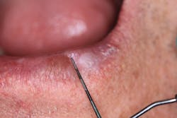
Upon performing a head, neck, and oral cancer screening, a 6 mm x 9 mm lesion was noted on the lower left side of the patient’s lip. The lesion was irregular in shape and white in appearance (figures 1 and 2). There was no sluffing or pain. Upon inquiry, the patient stated that the lesion appeared approximately two months ago and did not go away. He did not recall any trauma to the area either.
Differentials
- Seborrheic keratosis
- Discoid lupus erythematosus
- Atopic dermatitis
- Squamous cell carcinoma
- Actinic keratosis
Definitive diagnosis: Actinic keratosis
Treatment and discussion
The patient was referred to his dermatologist for a follow-up, where he had a brush and punch biopsy performed under local anesthetic. Definitive diagnosis was actinic keratosis. He has subsequently been under follow-up care with no complications.
Actinic keratosis is one of the most common lesions that develops on the skin, predominantly due to cumulative sun exposure. Other common risk factors include history of sun exposure, fair or medium skin, genetic disorders (xeroderma pigmentosum, albinism), use of tanning beds, advanced age, male sex, immunosuppression, infection with human papillomavirus, and arsenic exposure.1
Symptoms are varied and as follows2:
- Rough, dry, or scaly patch of skin, commonly less than 2.5 cm
- Flat to slightly raised patch or bump on the top layer of skin
- Sometimes a hard, wartlike surface
- Color variation—pink, red, brown, and white
- Itching or burning in affected area
Areas most exposed to the sun are the predominant locations for these lesions, which include scalp/neck, forearms, hands, ears, lips, and face.
Preventions include limiting time in the sun, covering up exposed areas, and liberal use of sunscreen.
Diagnosis is usually by a simple visual examination. However, a biopsy is recommended for a definitive diagnosis to assess the severity of some lesions, as these lesions will progress into squamous cell carcinoma.3
Complete removal of the lesion is common and ensures that it won’t return. Medication also can be applied in instances where several patches are found. A dermatologist will make the best recommendation on a case-by-case basis.
References
- O’Rourke KM. Haider A. Actinic keratosis: Diagnosis and treatment options. Medscape website. https://www.medscape.org/viewarticle/760020_2. Published July 31, 2012. Accessed July 8, 2019.
- Mayo Clinic Staff. Actinic keratosis. Symptoms & causes. Overview. Mayo Clinic website. https://www.mayoclinic.org/diseases-conditions/actinic-keratosis/symptoms-causes/syc-20354969. Published January 9, 2019. Accessed July 8, 2019.
- Mayo Clinic Staff. Actinic keratosis. Diagnosis & treatment. Diagnosis. Mayo Clinic website. https://www.mayoclinic.org/diseases-conditions/actinic-keratosis/diagnosis-treatment/drc-20354975. Published January 9, 2019. Accessed July 8, 2019.
About the Author
Stacey L. Gividen, DDS
Stacey L. Gividen, DDS, a graduate of Marquette University School of Dentistry, is in private practice in Montana. She is a guest lecturer at the University of Montana in the Anatomy and Physiology Department. Dr. Gividen has contributed to DentistryIQ, Perio-Implant Advisory, and Dental Economics. You may contact her at [email protected].


