Acute primary herpetic gingivostomatitis: A case report
Editor's note: Originally published December 15, 2014. Updated formatting August 19, 2022.
Lesions of the oral cavity manifest in a multitude of ways. Deciphering the information we observe and rendering a diagnosis and proper treatment can often be difficult due to resemblance in color, shape/size, and presentation similarity. This was the case for the situation described here.
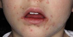
A healthy 2-year-old male presented to his general practitioner for a follow-up exam regarding some large, asymptomatic nodules on the right side of his neck. During the tonsilar/intraoral examination, a few red, ulcer-like lesions were noted on the buccal mucosa. Appetite and temperature were described as normal. The physician diagnosed the intraoral lesions as hand, foot and mouth disease (caused by the Coxsackie A and B viruses).
An extreme case of oral herpes
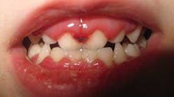
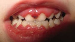
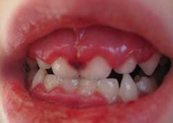
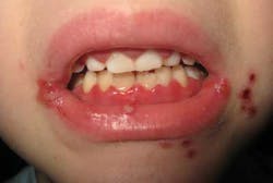
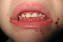
As the infection advanced, the gingiva became fire-engine red, very swollen, and tender to palpation. Any minute trauma to the papillae caused bleeding and severe pain. The attached gingiva and intraoral tissue became more inflamed, and oral hygiene was nearly impossible as the gums had practically covered the surfaces of the teeth. The lips became hemorrhagic and started crusting over.
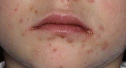
Later, the patient developed a mild fever with a widespread breakout of clusters of vesicles in the perioral area and the commissures of the lips. The crust-like nature of lesions in the oral and lip area made it very difficult to open and close the mouth. There was a fetid odor to the breath and an overall lethargic nature to the patient’s demeanor. Myalgia was present in the head and neck area. Eating was difficult, and the patient became quite irritable and slightly dehydrated.
Later, the patient developed a mild fever with a widespread breakout of clusters of vesicles in the perioral area and the commissures of the lips. The crust-like nature of lesions in the oral and lip area made it very difficult to open and close the mouth. There was a fetid odor to the breath and an overall lethargic nature to the patient’s demeanor. Myalgia was present in the head and neck area. Eating was difficult, and the patient became quite irritable and slightly dehydrated.
For the record, the patient had two older siblings who six days later began to manifest some of the same intra- and perioral vesicles. It is worthy to note that lesions were not observed on the hands or feet of any of the patients.
At this point, it was clear that the patient did not have hand, foot and mouth disease and was given the diagnosis of acute primary herpetic gingivostomatitis (APHG).
Herpes simplex virus (HSV) types 1 and 2 belong to the family Herpesviridae, which also includes Epstein-Barr, cytomegalovirus, and varicella-zoster viruses.1 Approximately 80% to 90% of the adult human population has been infected with HSV.1 Approximately 1% of initial oral infections occur as APHG2 as contact with the highly contagious secretions result in more than half a million cases reported annually in the United States.1 HSV-1 is the primary cause of APHG; however, HSV-2 can cause APHG by oral-genital or oral-oral contact. The primary infection usually affects children under the age of 10; a secondary/recurrent infection can occur in patients from 15 to 25 years of age. After a three- to 10-day incubation period, the infection starts to manifest with the aforementioned signs and symptoms. After 12 to 20 days, APHG will spontaneously regress.
Treatment is palliative. Liquids with caloric value should be administered due to the pain caused by the mouth ulcers and swelling of the gums. Prescriptions of the following can be given:
- Topical oral anesthetics—i.e., Orabase with benzocaine or lidocaine HCl 2% viscous solution
- Acyclovir ointment 5%
- Acyclovir 5%/lidocaine 2% compounded in a paste for perioral application
Lesions that coalesce in the perioral area must be monitored for secondary infections, especially if the patient is young and has a tendency not to leave the area alone.
References
1. Langlais RP, Miller CS. Color Atlas of Common Oral Diseases. Williams & Wilkins. 1992; 84,118.
2. Sapp JP, Eversole LR, Wysocki GP. Contemporary Oral and Maxillofacial Pathology. Mosby. 200-201.
About the Author
Stacey L. Gividen, DDS
Stacey L. Gividen, DDS, a graduate of Marquette University School of Dentistry, is in private practice in Montana. She is a guest lecturer at the University of Montana in the Anatomy and Physiology Department. Dr. Gividen has contributed to DentistryIQ, Perio-Implant Advisory, and Dental Economics. You may contact her at [email protected].
