Pathology case: Uncommon presentation with respect to location
Editor's note: Originally published June 26, 2018. Updated December 2024.
Presentation and patient history
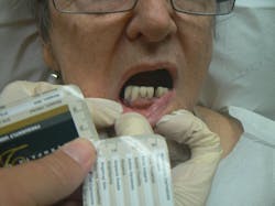
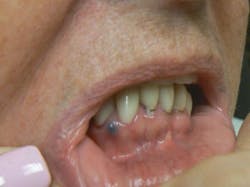
A 74-yr-old female presented with the complaint of an asymptomatic “bump” on her lower gums (figures 1 and 2). The patient stated that the lesion had been present for 10 months.
A previous general dentist had referred her to an endodontist who performed root canal treatment on her tooth. At that time, the endodontist lanced the lesion and a clear fluid came out. He then placed the patient on a prescription of the antibiotic amoxicillin. However, the papule did not disappear and became darker. The patient presented to a dermatologist for further evaluation.
Clinical exam
Physical examination revealed a 3 mm x 3 mm purple papule located on the lower attached gingiva between teeth nos. 27 and 28. Palpation revealed a soft, fluctuant nodule that was asymptomatic to the touch.
Differentials
- Amalgam tattoo
- Periodontal abscess
- Fibroma
- Pyogenic granuloma
- Gingival cyst
- Mucocele
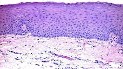
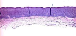
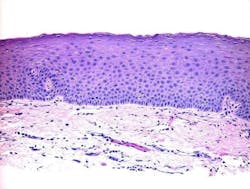
A pathology report was ordered, a deep-shave biopsy was performed, and the nodule removed. Pathology showed the mucus membrane with compressed fibrous tissue within the lamina proper and associated sparse inflammation (figures 3 and 4).
Final diagnosis
Mucocele or mucous retention cyst
Discussion
This report describes a lesion of the lower attached gingiva that was definitively diagnosed by histologic examination as a mucocele or mucous retention cyst. The usual location of mucoceles is on the lower lip. However, this case illustrates an uncommon presentation of a mucocele with respect to location.
Mucoceles are benign lesions that are filled with mucus and can appear in the mouth, appendix, paranasal sinuses, lacrimal sac, appendix, or gall bladder.1 Generally, mucoceles occur as a sphere and are asymptomatic. Mucocele is the seventeenth most common salivary gland lesion found in the oral cavity. The clinical appearance is typically a distinct, fluctuant, painless, bluish swelling of the mucosa. Most lesions are smaller than 1 cm in diameter. They form due to mucus accumulating when there is an alteration in the minor salivary gland that leads to limited swelling. Mucoceles typically do not form on the attached gingiva.
Mucoceles are cystic lesions that result from the accumulation of mucus due to an alteration in the minor salivary gland. This, in turn, causes swelling. The most common sites for them to occur are the lower lip, the tongue, the ranula (floor of the mouth), and the buccal mucosa.2
The diagnosis and management of a mucocele can be a challenging task. Surgical excision is the most common treatment with the least recurrence rate.
Here, we described an unusual case of a mucocele on the attached gingiva. A history of trauma to the mouth may be elicited to explain such an unusual presentation of the lesion; however, in this case, the patient did not recall any trauma. Clinical and histological findings confirmed the diagnosis. Although mucoceles are normally found in the first three decades of life,3 it was another oddity that this developed in our 74-year-old patient.
The close resemblance and clinical presentation of other possible lesions (amalgam tattoo, pyogenic granuloma, periodontal cyst, gingival cyst, etc.) pose a diagnostic challenge. Thus, excision and biopsy are warranted for definitive diagnosis and appropriate treatment.
References
- Regezi JA, Sciubba JJ. Salivary gland diseases. In: Oral pathology: clinical pathologic correlations. Philadelphia, PA: W.B. Saunders Co.; 1989:225-283.
- Senthilkumar B, Mahabob MN. Mucocele: An unusual presentation of the minor salivary gland lesion. J Pharm Bioallied Sci. 2012;4(suppl 2):180S-182S. doi:10.4103/0975-7406.100265
- Rao PK, Hegde D, Shetty SR, Chatra L, Shenai P. Oral mucocele—diagnosis and management. J Dent, Medicine and Medical Sci. 2012;2(2):26-30. https://www.interesjournals.org/articles/oral-mucocele--diagnosis-and-management.pdf
About the Author

Vincent J. Monticciolo, DDS, JD, MBA
Vincent J. Monticciolo, DDS, JD, MBA, is a graduate of the University of Detroit School of Dentistry. He earned his master's degree in business administration in 2001 and later his law degree in 2013. Early on, he found a way to improve his community by offering free dental care to struggling individuals who could not afford it. He and his wife, Dr. Natalie Monticciolo, began to think of how they could take this simple concept and turn it into something larger. After relocating to the Tampa, Florida, area, they formed Dentistry from the Heart in 2001. Today, the charity helps coordinate more than 250 events in all 50 states, as well as internationally. Dr. Monticciolo practices full time in one of his six dental offices near Tampa Bay.

Natalie Lama Monticciolo, DO
Natalie Lama Monticciolo, DO, is a board-certified dermatologist practicing in the Tampa Bay area in Florida. She founded and operates Prestige Dermatology. After receiving her medical degree from Michigan State University, she did her residency at NOVA Southeastern University. Her knowledge in skin diseases and cosmetic procedures comes from her extensive training, but her passion lies in skin cancer surgery. It is most rewarding to her when she helps others suffering from nonmelanoma skin cancer, melanoma, and other lesions that must be surgically removed.

Sophia Monticciolo
Sophia Monticciolo is a high school student with a keen interest in pursuing dentistry after college. While she excels in all of her classes, she thoroughly enjoys her science courses. In 2014, she, along with her sister, started a charity called Socks 4 Love. The socks are filled with daily necessities—such as toothbrushes, toothpaste, sunscreen, soap, and lotion—and delivered to the less fortunate in her community and as far away as Peru. More than 7,000 prefilled socks have been donated since the charity’s inception.
