5 tips to help intraoral exams serve as myofunctional airway screenings

Airway has been an evolving buzzword in dentistry for some time, and it’s not going anywhere. We already know the oral cavity is the gateway to overall health. We also know that we can survive three weeks without food and three days without water, but we can survive only three minutes without air. We don’t give much thought to our ability to breathe even though it’s essential to our overall well-being. In fact, it’s quite often an overlooked facet of our health.
Did you know that the maxilla is also the floor of the nose? As registered dental hygienists, we have the advantage to properly screen our patients for airway issues two or three times a year, sometimes more. In 2017, the American Dental Association released a policy statement encouraging dental professionals to screen for red flags due to airway obstruction. Let’s walk through an intraoral exam and I’ll show you how it can double as a myofunctional airway screener.
Begin your screening before you lay the patient back in the chair. As you’re chatting and reviewing their medical history, observe them. How are they breathing—through their nose or mouth? Where do you see the primary movement coming from—the diaphragm or the chest? Are their lips closed? We’re looking for dominant nasal breathing patterns with primary movement from the diaphragm and optimal oral rest posture, which includes the lips closed, the entire tongue lightly suctioned in the roof of the mouth, and the posterior teeth 2-3 mm apart.
You might also be interested in
BROOMS: Systematic screening for oral myofunctional disorders
Airway Revolution: “A mission to help millions”
3 hidden benefits of clear aligner therapy relating to airway health
Oropharyngeal area
Tell your patient you’re performing an airway assessment. Mallampati score should be performed while the patient is in a neutral position—ask them to open and extend their tongue. It’s important during assessment that the patient is not saying “ah” as this could cause a false reading. Essentially, you’re looking to see what structures are visible.
- Class 1: soft palate, fauces, uvula, anterior and posterior pillars visible
- Class 2: soft palate, fauces, uvula visible
- Class 3: soft palate, base of uvula visible
- Class 4: soft palate not visible
Class 3 and Class 4 Mallampati scores are indicative that a patient has a narrow airway and increased chance of having obstructive sleep apnea.1 I recommend printing and laminating a photo of the Mallampati score; you may find that patients ask for more information before you even begin.
Once the patient is laid back, you’re already examining the oropharyngeal area for any abnormalities. This is a great time to examine and grade the patient’s tonsils. Enlarged tonsils, grade 3 or 4, warrant a referral to an otolaryngologist as the tonsils may be causing airway obstruction.
- Grade 1: tonsils occupy <25% of the oropharynx
- Grade 2: tonsils occupy 26%-50% of the oropharynx
- Grade 3: tonsils occupy 51%-75% of the oropharynx
- Grade 4: tonsils occupy >75% of the oropharynx
Hard palate
Next, examine the hard palate. Aside from your routine protocol, look at the size and shape of the maxilla, along with any dental crowding. To measure the transverse width, take a cotton roll and hold it up between tooth nos. 3 and 14. Cotton rolls are generally ~36-37 mm. According to Dr. James A. McNamara, a range of 36 to 39 mm indicates a maxillary arch that can accommodate a dentition without crowding or spacing.2 The maxilla is also the floor of the nose and the lateral walls of the nasal cavity. If we have insufficient maxillary development, we will also have insufficient oropharyngeal space.
I encourage you to look at palatal rugae in a different light. Prominent palatal rugae may indicate poor tongue rest posture. Pressure from the tongue resting in the roof of the mouth should flatten the palatal rugae. Low tongue rest posture may also obstruct the airway.
Tori are something we already look for as an atypical finding in our exams. However, did you know that tori are associated with bruxism? Yet bruxism may still be present without the finding of tori. Even though bruxism would be noted during your hard structure assessment, there’s a connection between bruxism and airway.
According to sleep medicine specialist Jerald Simmons, MD, and sleep dental specialist Ronald Prehn, DDS, “When most patients exhibit obstructive respirations during sleep, the mandible falls back, bringing the back of the tongue with it. This triggers a series of events that in some people results in a reflexive attempt to open the airway by increasing masseter tone. This brings the mandible forward and, in many patients, improves respirations. We postulate [sic] that nocturnal bruxism is a compensatory mechanism of the upper airway to help overcome upper airway obstruction by activation of the clenching muscles, which results in bringing the mandible, and therefore the tongue, forward.”3
Labial mucosa
A good time to examine the lips at rest is when palpating the labial mucosa. Do you notice any strain? The mentalis muscle may over activate in an attempt to close the lips. This gives a stippling appearance in the muscle. Dry and cracked lips may indicate mouth breathing.
The tongue
We often ask the patient to raise their tongue to the roof of the mouth when we examine the ventral surface of the tongue. This is a good time to assess the range of motion of the lingual frenum. Are they able to reach the roof of their mouth, or do they need to close their jaw to do so? If a restricted lingual frenum is in question, a functional evaluation is warranted by an orofacial myologist. Simply asking a patient to stick out their tongue does not suffice.
In dental hygiene school, we’re taught to evaluate ankyloglossia by seeing if a patient can extend their tongue tip past their lower lip. Protrusion is one of the many functions of the tongue, which should be viewed as a muscular and respiratory organ. It’s involved in optimal craniofacial respiratory growth and development, mastication, swallowing, speaking, and keeping the upper airway open during sleep. Tongue scalloping is associated with a higher Mallampati score and should prompt questions regarding sleep and snoring history.4
Floor of the mouth
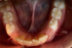
A good time to see where the lingual frenum attaches is when you examine the floor of the mouth. Does it attach into the floor of the mouth as it should? Or do you see it attaching to the alveolar ridge? When this attachment is present, it often presents as an “Eiffel tower” appearance (figure 1). This appearance may be indicative of a posterior lingual restriction. Further functional evaluation is warranted by an orofacial myologist.
I challenge you to continue expanding your role as a registered dental hygienist. I truly believe we play an integral role in identifying key signs of airway obstruction. I hope after reading this you feel empowered to add these critical steps to your intraoral evaluation. We have the ability to impact our patients’ lives outside the dental operatory in ways we never imagined. You’re already there; I’m just giving you a new lens to see through. Dig deeper, look for root causes of disease, and remember, airway trumps all.
References
- O’Brien SM. Understanding the Mallampati score. Clinical Advisor. February 4, 2016. http://www.clinicaladvisor.com/home/the-waiting-room/understanding-the-mallampati-score
- McNamara JA. Maxillary transverse deficiency. Am J Orthod Dentofac Orthop. 2000;117(5):567-570. doi:10.1016/S0889-5406(00)70202-2
- Simmons J, Prehn R. Nocturnal bruxism as a protective mechanism against obstructive breathing during sleep. Accessed December 19, 2021. https://csma.clinic/Bruxism_Poster.pdf
- Weiss TM, Atanasov S, Calhoun KH. The association of tongue scalloping with obstructive sleep apnea and related sleep pathology. Otolaryngol Head Neck Surg. 2005;133(6):966-971. doi:10.1016/j.otohns.2005.07.018
About the Author
Brittny Murphy, BS, RDH, MAS, COM,
Brittny Murphy, BS, RDH, MAS, COM, is a registered dental hygienist, myofunctional therapist, educator, author, and key opinion leader in sleep and myofunctional therapy. She is a Buteyko breathing educator. Brittny is the founder of CT Orofacial Myology, a private myofunctional therapy practice aimed at improving oral and whole-body wellness, through which she has helped hundreds of patients thrive by sleeping and breathing better. Brittny is also the face behind the podcast, “I Spy with My Myo Eye.”