Tools to relieve patient anxiety
Technology provides new approaches
By Mary Govoni, CDA, RDA, RDH, MBA
When I ask dental teams about their most important goal in treating patients, they frequently respond with statements about making dentistry comfortable and less frightening. To that end, we have many tools and techniques available to help patients overcome anxiety about dental treatment. Developing the right verbal skills to reassure them about minimizing discomfort, describing the various steps in each procedure, medication, and hypnosis are some examples of techniques that many dental team members utilize. With spa dentistry becoming more popular, massage and aromatherapy also are effective methods of increasing patient comfort. Noise reduction headsets, virtual reality glasses, video programming, and CD players with relaxing music are some examples of technology that might be employed with apprehensive patients.
While all of these techniques might be effective, they serve to distract patients or mask their underlying cause of anxiety - fear. If we as dental professionals address patients’ underlying fears about dental procedures, we can greatly reduce their anxiety and perhaps make dentistry an enjoyable experience. It’s been said that the greatest fear is that of the unknown. The best way to eliminate this fear is to make the unknown known.
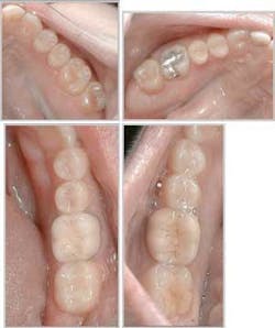
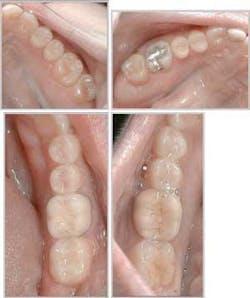
To make the “unknowns” about dental treatment known to patients, we must educate patients. No big surprise here - we strive to educate our patients every day. Yet many of them still feel anxious about treatment. Imaging technology can provide some powerful tools to enhance that education, and eliminate the unknowns. Remember the old saying, “A picture is worth a thousand words”? It is so true for dentistry. Not just any picture will do, however. The picture needs to be that of a patient’s own mouth, and must show existing conditions. Digital radiography and intraoral photography serve well to produce powerful real-time images that help patients understand their need for treatment. These images also can provide reinforcement for patients who desire optimum oral health and help them translate their needs into wants. Patients can more readily see decay, fractures, or bone loss on a full screen view of a radiograph rather than on a 1.25” X 1” traditional film. Teeth and surrounding structures can be viewed with greater ease (and impact) on digital images or videos displayed on a computer screen than by a patient looking at his or her mouth in a hand mirror. In addition, digital radiographs and images can be magnified - and certain enhancements made to the image - in order to focus on a specific area of concern.
For a new and/or emergency patient, a video clip or “tour” of the individual’s mouth is an excellent way to introduce the diagnostic exam. It also can be used in presenting a treatment plan. Intraoral cameras have the capability to capture short video clips that can be stored in a patient’s electronic chart in most practice-management software programs. The camera moves slowly across both arches and can stop or zoom in on particular areas of interest. Copies of this video clip, or any still images, can be burned to a CD for a patient to take home.
Still images can be captured with an intraoral camera or with a digital still camera. The images should be captured in a particular order to establish a routine and increase efficiency. This is very similar to exposing a complete mouth series of radiographs. Starting in the same area of the mouth and following a system for exposures help to ensure that all necessary views are captured. Dr. John Jameson of Jameson Management, Inc. recommends the following images be taken - a full face view, a smile view, a retracted smile view, a view with the cheeks retracted and the mouth slightly open, buccal views of both right and left sides of the mouth, occlusal views of the upper and lower arches or separate views of each of the four quadrants, lower anterior linguals (due to typically high calculus build-up), and any problem areas.
It may take some practice to become proficient at capturing these images, but the investment in training and practice will be well worth it. Not only can this type of education help patients feel less anxious about their care, their acceptance of necessary treatment will most likely increase.
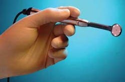
This is just one example of how technology can educate a patient and allay fears prior to treatment. But what about during treatment? Intraoral cameras and imaging devices come to the rescue again. Using an intraoral camera during some phases of treatment can help to reassure a patient about what is happening in his or her mouth. This returns us to the fear of the unknown. A new tool, which combines the use of an imaging device and a mouth mirror, is now available for use during examination and treatment. The Mirroscope™ from Miras Mirror Imaging Solutions allows the dentist, assistant, or hygienist to use the mouth mirror to see the structures in the mouth while those images are transferred in streaming real-time video to a computer monitor. This, in turn, can be viewed by a patient. The technology also is designed to allow dental professionals to work from a video monitor, rather than the mouth, in a magnified view that is comparable to a medical procedure performed with a laparoscope. Utilizing this new technology requires practice to develop skill and efficiency. But, once again, it appears worth the investment.
Using imaging technology is not the ultimate solution to treating anxious patients. But it certainly can enhance patients’ experiences, increase their knowledge about oral health, and assist them in making better choices about their treatment. Sounds like a “win-win” to me.
Mary Govoni is a Certified and Registered Dental Assistant and a Registered Dental Hygienist with more than 28 years of experience in the dental profession as a chairside assistant, office administrator, clinical hygienist, educator, consultant, and speaker. She is the owner of Clinical Dynamics, a consulting company dedicated to the enhancement of the clinical and communication skills of dental teams. She can be reached at [email protected].


