High-tech simulators come to the preclinical lab
How many of us remember our preclinical experience? We learned cavity preparations with typodonts on a pole, on benchtop, or if we were lucky, maybe even on a manikin.
We have truly transcended into the 21st century, using a system that can be compared to guided missile technology!
As the use of technology has evolved in clinical dentistry, so has it been implemented in dental education. One of the newest, most exciting tools available is the Virtual Reality Dental Simulation system (VRDS). This educational instrument helps the student develop early psychomotor training in tooth preparation for direct and indirect restorations.
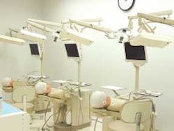
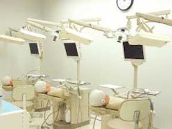
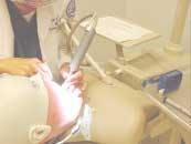
The VRDS consists of a simulator unit connected to a computer system. The system described in this article is the DentSim™, manufactured by DenX, Ltd.1 The simulator unit includes a typodont and manikin head attached to a simulated patient's chair and torso. It is equipped with a standard overhead light, handpiece attachments, rheostat, air/water syringe, and suction. The computer system consists of a CPU with installed software, high-speed handpiece with LED sensors, typodont with a reference body LED sensor attached to it, and an infrared light camera. There is also a flat-screen monitor and a mouse (Figure 1). The operator (e.g., student) accesses the system and reviews the parameters of the assigned tooth preparation before beginning the clinical procedure. An additional software feature provides tutorials about related topics.
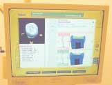
As the operator/student performs an assigned tooth preparation, the camera reads the position of the handpiece in relation to the typodont tooth. This information is simultaneously transferred to the database. The user is then able to immediately compare his or her performance with the preprogrammed "optimal" tooth preparation (Figure 2). An example of the evaluation criteria includes outline form, removal of carious lesions, floor depth, damage to adjacent teeth, smoothness of preparation, retention form, and many other detailed criteria. For instance, if a student performs a clinical procedure and prepares the cavity too deep, the computer will emit a "ding, ding, ding" sound to let the student know that the pulp is exposed. At the completion of any procedure, the operator may use a "playback" menu, reviewing all of the work in graphic video mode. Students generally adapt very easily to this system, since they are familiar with interactive computer technology.
Several disciplines of dentistry are already included in this system, such as operative dentistry, fixed prosthodontics, aesthetic dentistry, and endodontics. More specifically, the procedures available are tooth preparations for amalgam and composite resin; preparations for inlay, onlay, full gold crown, porcelain-fused-to-metal, and all-ceramic crown; and endodontic access. Other programs are being developed and will be available soon. The faculty can customize each preparation according to the philosophy of the dental institution implementing the system. In this way, use of VRDS can be incorporated into an ongoing dental curriculum.
VRDS at Nova Southeastern University
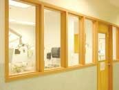
At Nova Southeastern University's College of Dental Medicine (NSU-CDM), the VRDS was introduced in September 2003. The Virtual Reality Laboratory includes six units and is centrally located near faculty offices and other research laboratories. Students, faculty, and administrators pass by this area regularly and are able to observe the units in use (Figure 3). Entering freshmen are assigned an equal number of rotations in which they are given orientation and assigned specific projects.
Advantages of VRDS
We have observed several advantages of this early exposure to VRDS at Nova:
- Reinforcement of concurrently learned dental concepts — Students can apply biologically sound concepts from the freshman dental anatomy and cariology courses directly to a simulated clinical experience.
- Correct ergonomic positioning — Incorrect operator and/or patient positioning can result in blocking the camera from reading the LED sensors. When this happens, a warning signal prevents the user from continuing. Accommodations are also available for left-handed students (Figure 4).
- Correct use of dental instruments — Students learn how to correctly use the high-speed handpiece, rotary cutting instruments, mirror, explorer, and periodontal probe during the first semester of dental school.
- Psychomotor skills — Training in direct and indirect vision and spatial orientation in a timed setting are incorporated very early into the dental curriculum with VRDS.
- Self-evaluation — Students have unlimited, immediate, and objective access to detailed feedback of their work. The imaging available in this system includes 3-D graphics, cross-sections, measurements, and zoom features. With VRDS, students may view their procedures from a 3-dimensional perspective.
- Faster acquisition of skills — The research that has been published so far indicates that students attain a competency-based skill level at a faster rate than with traditional simulator units.2 This can result in a domino effect; i.e., changes in dental curriculum, course sequencing, and perhaps earlier entrance into the predoctoral clinic.
- Positive student perception — The majority of courses in the freshman curriculum relate to basic biomedical sciences. Students seem to enjoy the opportunity to have what they perceive as a more dental-related course with VRDS.
Disadvantages of VRDS
As with any system, several disadvantages with VRDS do exist:
- System limitations — The system is programmed for and evaluates tooth preparations only. Tooth restoration is not included in the program at this time. In addition, hand instrumentation cannot be used.
- Cost — Units are expensive. The cost of each unit is approximately $70,000.1 New programs are also expensive at about $6,000 each.
- Equipment maintenance and repair — Technology-based systems require faculty/staff to be available for training, repair, and supervision of the laboratory. The system is made up of components that come from more than one manufacturer. The manikin head, typodont, and computer system may come from three different manufacturers. Troubleshooting and equipment repair are not difficult, but sometimes must be handled through all of the companies involved.
Clinical uses of virtual reality
The most direct clinical utilization of this program exists in the IGI™ system, also by DenX, Ltd.1 This system is used in clinical dentistry for Image Guided Implant placement. In this system, the jaw CT scan of the patient is input into the computer database. The parameters of correct implant placement are then applied in 3-D to the patient's CT scan. The implant is then placed in correlation to this 3-D image. The technology used in dental education has been broadened for direct application in clinical dentistry. Implant placement continues to require precision and knowledge on the part of the dentist. The image-guided system improves visualization for the dentist and education for the patient.
The potential for use of this system is enormous. Research in dental education related to teaching methodologies, psychomotor skills, and faculty standardization are all possible.
Collaboration research among the universities using this system is in the development stage. The participating institutions involved in this collaborative effort are known as the Advanced Simulation Research Group.3 The system provides an additional learning option for the student, as well as standardization of procedures and objective grading systems for the faculty. Some of the future possible applications for the dental clinician include regional board examinations, recertification examinations, and continuing-education courses utilizing this technology.
Virtual reality systems have been in use less than 10 years, and currently are being used in about a dozen dental institutions. There is still much to be learned about this teaching method. Nevertheless, the future is here, and we all should take advantage of the capabilities of this exciting new technology.
The authors would like to acknowledge Dr. Harvey Quinton, assistant professor, Department of Restorative Dentistry, for his collaboration in the NSU Virtual Reality teaching laboratory.
References
- DenX, Ltd., Melbourne, Australia. www.denX.com.
- Urbankova A, Lichtenthal R. DentSim virtual reality in preclinical operative dentistry to improve psychomotor skill — a pilot study. J Dent Educ 2002; 66(2):284.
- DentSim Research Collaboration Meeting, University of Pennsylvania, School of Dental Medicine. June 10-11, 2003.
Abby J. Brodie, DMD
Dr. Brodie is an associate professor in the Department of Restorative Dentistry at Nova Southeastern University, College of Dental Medicine, Fort Lauderdale, Fla. Her professional focus is preclinical education and dental curriculum. Contact Dr. Brodie at [email protected].
Judith L. Gartner, DMD, MMSc
Dr. Gartner is an assistant professor in the Department of Prosthodontics at Nova Southeastern University, College of Dental Medicine, Fort Lauderdale, Fla. She is also in private practice focusing on aesthetic dentistry and prosthodontics in North Miami Beach, Fla. Contact Dr. Gartner at [email protected].
