No more supremely stinky teeth: using 3-D printed replicas to teach endodontic techniques
Extracted human teeth have been a blessing and a curse for endodontic educators.
The blessing? Teaching dental students and dentists new endo techniques in real human teeth without the human attached. Awesome, no human! The curse? Infected, supremely stinky teeth with unknown anatomy, no relationship to educational objectives, and no mulligans.
Let’s put aside the supremely stinky teeth part for now and just look at the unknown anatomy issue, the educational objectives issue, the testing issue, and the no mulligans issue.
The unknown anatomy issue doesn’t need much explanation. Unknown anatomy is the fun part of clinical endodontic practice, but it’s a nightmare in procedural endodontic training. Think about it. Teaching endo technique is limited by the random nature of the root canal anatomy presented in the extracted teeth students procure. It’s not like you get to choose the anatomic challenges that your students work their way through, or ever know what’s in those teeth they brought to your course.
ADDITIONAL READING |Saving teeth through surgical endodontics
The educational objectives issue is very simple. There are a number of procedural endodontic challenges — abrupt apical curves, “S” curves, cervical curves, mid-root bifurcations, apical confluencies, etc. How do you find a number of extracted teeth with the same anatomic challenge you want to teach your students to handle? Not possible with extracted teeth; they aren’t designed to answer a specific procedural training objective.
ADDITIONAL READING |The perio-endo overlap
The testing issue? Extracted teeth for board exams are neither fair to the examined nor defendable as fair by examiners. No consistency, no defense, case closed.
The no mulligans issue? Being denied another attempt after you’ve just screwed the pooch working through a given anatomic challenge? That’s rough, and that’s why becoming competent in endo therapy has been a random walk through endo anatomy only overcome by a large number of cases treated.
Up until now, endodontic procedural training has been hamstrung by the random nature of the anatomy encountered by students when working in extracted teeth (not to mention the smell). When we attempt a procedure in a given root canal space and, for instance, come upon an impediment in the canal but ledge it before we know better, we don’t get a second attempt. We are left to hopefully deconstruct the error correctly, and then just wait for another tooth having similar anatomy.
So what would it be like to repeat — in exactly the same anatomic form — a procedure over and over (and over, if necessary), until you get it?
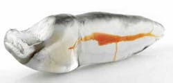
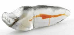
Fig. 1: TrueTooth™ procedural training replica. Note the fine detail of internal root canal anatomy, including fins, webs, and loops, lateral and accessory canals — the full Monty.
Flash back to more than 20 years ago …
I treat a physics professor from UCSB who explains how he invented a method to print, in three dimensions, any computer-generated CAD model — a process he called stereo-lithography. The machine he invented had a 10-inch cubed vat — with a plate that moved up and down in it — filled with photo-polymerizing resin. Above that a laser traces the surface of the solution, polymerizing, layer-by-layer, the cross-sectional geometry of the object to be fabricated. The plate then drops by a precise amount, and the laser traces the next layer. When the object has been completely printed, the plate rises out of the resin bath with the object revealed.
This blew my mind completely. Everything was different now. Research, prototyping, testing, training, manufacturing, engineering, product design and pretty much everything else was, or was going to be, different now. My first question?
Could we print replicas of teeth?
I had spent the previous five years working out how to CT scan extracted teeth and reconstruct them in 3-D computer space as virtual replicas, (1) so replicating, in 3-D, the incredible anatomy we were seeing in our reconstructed models was very fun to consider.
He said no, not yet — the technology was in early development so the resolution was too large to work for RCT training and the cost of printing at that point could only be borne by aerospace firms. So I waited for 10 years, and had a tooth replica printed at the resolution possible at that time but found it was not yet ready for prime time in dentistry (Fig. 1). Last year, the inflection points of resolution, polymer chemistry, and cost intersected for dentistry’s use of stereo-lithography, and this year Dental Education Laboratories, after completing our development, introduced as TrueTooth™ training replicas.
It’s here now
These replicas are printed in clear and opaque versions for training and testing, and the polymers used are heat-resistant, so high-speed burs don’t gum up when cutting access cavities and the replica’s canal walls don’t melt when obturating with warm gutta-percha methods. Inside the replicas, in all the root canal spaces, is a gel-like material that is red in color and dissolves in sodium hypochlorite, immediately showing students the efficacy of their irrigation procedures. Inside the replicas are the phenomenal intricacies of endodontic anatomy in all of its natural glory.
Negotiating through the canals gives tactile feedback that there is still “pulp” medium present, that the file tip has engaged a lateral fin or accessory canal — in other words, almost as good as the real thing. Cutting shapes in clear TrueTooth roots reveals what aggressive file tips are actually doing when they destroy apical anatomy during preparation procedures. Obturation procedures in these replicas are a “thrill of the fill” experience.
Summary? All boxes are ticked:
- No unknown anatomy. All the anatomy of every replica is known.
- Replicas can be chosen for teaching specific endodontic procedural skills.
- Replicas are exactly the same so they are a fair test between board applicants.
- 3-D printed replicas can be worked with — over and over — the same challenge every time.
- They are less expensive than the oversimplified training models available.
- No more stinky teeth.
Endodontic procedural training will never be the same.
Reference
1. Buchanan LS, Hebert JP. 3-D computer reconstructions of CT scans of extracted human teeth. Unpublished, 1987.


