Teeth staining summary by color and origin for the dental hygiene exams
Review this summary to successfully pass the dental hygiene exams
By Claire Jeong, BS, MS, RDH
We have all probably tried to drink our coffee and even wine through a straw to avoid staining our teeth, but what else can cause stains? And can stains be removed? Let’s discover how we can use the color and pattern of stains to see how they occurred and whether they can be removed. If you are taking the dental hygiene board exams, try your best to memorize the summary of teeth staining below.
Staining by Color
The color of the stain tells a lot about how the discoloration occurred:
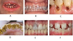
- Orange-red: chromogenic bacteria, chromic acid/copper chemicals
- Yellow: heavy dental biofilm/calculus
- Green: Nasmyth’s membrane(thin tissue on newly erupted teeth), copper/nickel chemicals
- Blue: dentinogenesis imperfecta, dentin dysplasia
- Brown spot: potential carious lesions, fluorosis
- Brown: chlorhexidine, tobacco, food (e.g., red wine, tea, coffee), stannous fluoride
- Black: iron/silver/manganese chemicals, betel leaves
- Grey: pulp necrosis, amalgam restoration
- White: demineralization, fluorosis
- Bands of dark colors: tetracycline
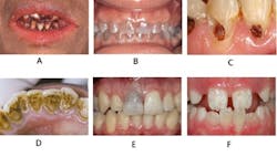
Figure caption:A - Betel leaf, B. Dentinogenesis imperfecta, C. Carious lesion, D. Tobacco, E. Pulp necrosis, F. Fluorosis
If you memorize this, you are ahead of the game. Now let’s expand on the topic of teeth discoloration by reviewing some definitions.
Exogenous vs Endogenous: When Did Staining Occur?
Did the discoloration occur during or after tooth development? There are two definitions related to “when” the staining occurred.
• Endogenous stain: occurred during tooth development
Fluorosis is considered an endogenous stain because the fluoride mineral affected the teeth while they were forming. Depending on the severity, affected teeth can have white/brown spots, milky opalescence, pitting, and also mottling. Tetracycline stain is also considered endogenous because the antibiotic consumed by the mother influenced the tooth development of the fetus.
• Exogenous stain: occurred after tooth eruption
Dietary pigments from foods and beverages are one of the most common causes of teeth staining. Pulpal necrosis, and its dark discoloration, is also considered a type of exogenous stain, if occurred after the tooth is completely formed and erupted.
Intrinsic vs Extrinsic: Where Is the Stain Located?
Is the stain outside or inside the tooth structure? There are two definitions related to “where” the stain is located.
• Intrinsic stain: located within the tooth
This type of stain cannot be removed by scaling and/or polishing. It may be reduced by whitening to some degree, but will not disappear easily. Examples of intrinsic stains include fluorosis, tetracycline stain, and pulpitis.
• Extrinsic stain: located on the surface of the tooth
Since the stain is on the outside of the tooth, it can be removed by scaling and/or polishing. But over time extrinsic stain can become intrinsic. Examples of extrinsic stains include food (e.g., coffee), tobacco, and plaque.
Review the example below.
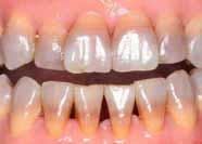
The discoloration shown in the photo is most likely caused by:
- Tetracycline
- Fluorosis
- Pulpal necrosis
- Tobacco
Answer: 1. Tetracycline
The teeth are yellow/brown/grey and have visible “bands.” Bands of discoloration are characteristics of severe tetracycline staining caused during teeth formation. For this reason, tetracycline is contraindicated for pregnant or lactating mothers. Fluorosis can create white/brown spots, milky opalescence, pitting, and also mottling, but is not characterized by bands of discoloration. Pulpal necrosis can cause darkening of the affected tooth (not the entire dentition). Tobacco can cause dark, irregular stains especially on the lingual surfaces of teeth.
In closing, try to apply this knowledge to case studies. See if the stains are related to medications (e.g., chlorhexidine) or habits (e.g., smoking) the patient may have. Lastly, go one step further to understand if the stain can be removed with scaling or polishing. Studies have shown that it takes at least 3 reviews to memorize new information. Ensure you continuously review this summary to successfully pass the dental hygiene exams and become the star clinician!
References
- Wilkins EM. (2013) Clinical Practice of the Dental Hygienist 11th ed. Philadelphia PA: Wolters Kluwer Health, Lippincott Williams & Wilkins.
- DeLong L, Burkhart NW. (2013) General and Oral Pathology for the Dental Hygienist. 2nd ed. Baltimore MD: Wolters Kluwer Health, Lippincott Williams & Wilkins.
- Ibsen O, Phelan JA. (2014) Oral Pathology for the Dental Hygienist. 6th ed. St. Louis, MO: Elsevier Saunders.
Claire Jeong is the founder of StudentRDH, a review solution for the National Dental Hygiene Board Exams, Local Anesthesia Board exams and CSCE. She graduated from MCPHS University, Forsyth School of Dental Hygiene and is a member of Sigma Phi Alpha, the dental hygiene honor society. Claire mentors students and provide speeches at dental hygiene programs related to the topic of dental hygiene board examinations. Claire is licensed in the United States and Canada. She provides personalized mentorship at StudentRDH and can be reached at [email protected].


