Determining treatment success in sleep apnea patients using tongue measurements
By Whitney Mostafiz © Dreamstime
According to new research that received the Graduate Student Research Award last month at the 19th Annual Meeting of the American Academy of Dental Sleep Medicine, measuring the ratio between tongue volume and bony enclosure size in patients with obstructive sleep apnea (OSA) may help dentists estimate oral appliance treatment success.
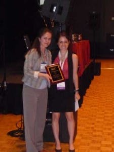
Whitney Mostafiz receiving the Graduate Student Research Award from Fernanda Almeida, DDS, on Saturday, June 5, 2010, at the Henry B. Gonzalez Convention Center in San Antonio, Texas.Using a mandibular advancement splint (MAS) has been shown to be a minimally invasive and effective treatment for OSA, which is an alternative to continuous positive airway pressure (CPAP). However, predicting efficacy of MAS treatment in individual patients is challenging. Through a collaboration between Sydney University and Harvard School of Dental Medicine, researchers assessed whether anatomical factors were associated with MAS treatment outcome. Some of these factors included craniofacial size, upper-airway soft tissue volume, and/or the anatomical balance between them. Cephalometric radiographs were digitized and craniofacial parameters were analyzed using Dolphin software, while the oral cavity area measurements were measured with ImageJ software. Dental cast measurements were used to calculate horizontal, vertical, and sagittal dimensions of the dentition as well.The study included 49 OSA patients. Patients were at least 18 years old and had mild to severe sleep apnea. They were without other sleep disorders or serious comorbid medical or psychiatric disorders. Each patient was fitted for a custom two-piece MAS, which was worn during sleep. Treatment outcome was assessed by polysomnography after approximately six weeks of oral appliance therapy. Of the 49 patients, 24 responded to the treatment and demonstrated an apnea-hypopnea index (AHI) reduction of 50% or more. Body mass index and age did not differ between responders and non-responders, but responders did have a lower baseline AHI, indicating that their sleep apnea was less severe before treatment. Tongue cross-sectional area (CSA) was measured in a subset of 28 patients, including 12 responders and 16 non-responders. The measurements were taken using cephalometric soft-tissue image analysis. Responders had a larger tongue CSA than non-responders, but there was no difference in the bony oral enclosure CSA. The ratio of tongue to bony enclosure CSA significantly differed between responders and non-responders, indicating the ratio as a significant predictor of response to treatment. Because patients who responded to MAS treatment had a larger tongue volume for a given oral cavity size, the researchers suggest that determining this ratio may help predict MAS treatment success. Nevertheless, imaging and the relationship between tongue and oral cavity volumes deem further exploration. Soft-tissue analysis through cephalometric imaging is more difficult to standardize, which ultimately resulted in a smaller sample size. CT imaging techniques may be useful for future tongue analysis as well.While this study reaffirms the difficulties in predicting OSA treatment response to mandibular advancement splints, responders seem to have a larger tongue volume for a given oral cavity site, suggesting that MAS may help correct anatomical imbalances. In the clinical setting, these measurements may be a practical tool in the treatment planning of OSA patients.Whitney Mostafiz is a third-year dental student at the Harvard School of Dental Medicine, and received a bachelor’s in science in human biology from Brown University in 2008. Her research interests include both sleep medicine and dentistry. Whitney has been involved in studies that have examined sleep deprivation in adolescents, dental imaging techniques, diet and tooth morphology in minnow species, and sleep apnea oral appliance therapy.


