The importance of piezo electric ultrasonics
By Kenneth Koch, DMD, and Dennis Brave, DDS
As followers of Real World Endo know, we are strong proponents of ultrasonics in dentistry. In fact, we believe the use of piezo electric ultrasonics is the most under-utilized technology in modern endodontics. However, we expect this to change in the very near future.
We prefer piezo electric ultrasonics - as compared to earlier magnostrictive units - for two reasons:
- Piezo electric technology offers more “cycles” per second (40,000 CPS versus 24,000 CPS).
- The tips of these units work in a linear, back and forth “piston-like” effect. This motion is ideal for endodontics. This is particularly evident when “troughing” for hidden canals. A magnostrictive unit, on the other hand, creates more of a figure 8 (elliptical) motion. This is not as ideal for surgical or non-surgical endodontic use.
Many dentists continue to have the impression that ultrasonic use in endodontics only has a surgical application. However, probably 90 percent of all ultrasonic use is in non-surgical endodontics. In our opinion, if you are serious about doing quality endodontics, you need to have a piezo electric ultrasonic.
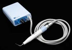
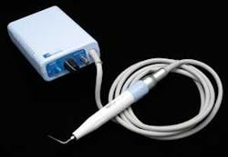
Once we grasp the notion of ultrasonics in endodontics, the next area of focus is the unit itself. This is where significant change has occurred. As part of our “EndoSequence System,” Real World Endo is proud to introduce a new ultrasonic - the “Varios 350.” The “Varios 350” (seen at right) is manufactured by NSK and distributed by Brasseler USA. Let’s take a closer look at this unit and some of its features.
The “Varios 350” is a piezo electric ultrasonic that fits in the palm of your hand! Yes, you have read the previous statement correctly. The 350 is the smallest unit on the market and it fits easily on any bracket table. It is so small that you can even place Velcro straps around it and place it anywhere. In fact, this unit (in combination with a portable handpiece) makes hygiene, as well as endodontics, truly portable. Additionally, the design of the unit allows an easy wipe down (disinfection).
While the size distinguishes this unit from all others, there is another feature that we think is the new dimension. The “Varios 350” comes with a fiber optic that is built into the handpiece. The brilliance of this design is that the fiber optic is protected and precisely directed to the field of treatment. This is unlike any other fiber optic because the light is completely circumferential and surrounds the tip. But why do we believe this is a quantum leap forward in ultrasonics? Let’s think, for a moment, about the benefits of fiber optics.
The primary benefit of having a fiber optic built into an ultrasonic is enhanced vision. Not only will clinicians see better, but the fiber optic actually creates a “wand of diagnostic ability” for the hygienist and dentist. By employing transillumination (shine the light through the tooth at the CEJ), we can now readily diagnose cracks, fractures, and even calcified canals. Additionally, think how great a fiber optic-based ultrasonic is to see subgingival calculus! For those clinicians performing endodontic or periodontal surgical procedures, the “Varios 350” will specifically illuminate your surgical site and make your life a lot easier.
The handpiece itself is small, light, and ergonomically designed for excellence. A thin handpiece, when combined with a fiber optic and the use of ultrasonic tips, allows the practitioner more visual access to the procedure. In endodontics, we have access to the tooth and straightline access to the canals, but we also need visual access to the procedure. Take a look at the “Varios 350” and pick up the handpiece. Take it for a “test drive” on a tooth. You will be stunned that such a small ultrasonic can be so powerful. We think you’ll like it!
Another critical factor in the use of piezo electric ultrasonics is the selection of tips. The “Varios 350” comes with a huge assortment of tips. Tips are available for endodontic, periodontal, and even general procedures. Additionally, Real World Endo is currently designing multiple new endodontic tips for both surgical and nonsurgical use. To further facilitate the use of this unit, we have created combination packs that include both endodontic (three) and periodontal tips (three). An ultrasonic tip caddy (UTC) helps keep things neat and organized in the treatment room.
Having just discussed the merits of what we think is an exciting unit (Varios 350), let’s review some of the uses of piezo electric ultrasonics in endodontics.
Finding hidden canals
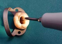
In addition to length control, the biggest challenge facing the general practitioner is finding the canals. Endodontic cases are becoming increasingly difficult, particularly those cases where the orifice has become occluded by secondary dentin. You can’t perform root canal therapy unless you find the orifice. Piezo electric ultrasonics are excellent for removing the secondary dentin that often slopes off the mesial wall. This is what blocks the MB-2 canal in maxillary molars. It is particularly in these maxillary molar cases, when looking for the MB-2, that the “Varios 350” shines! A good tip to remember when searching for hidden canals is that secondary dentin is generally whitish or opaque, while the floor of the chamber is darker and more gray-like in composition. The fiber optic can really help the clinician in these cases by making this an obvious distinction.
Piezo electric ultrasonics also perform particularly well when breaking through the calcification that covers the canal orifice. A troughing tip is a good choice for this task. As a result of the “graying of America,” along with the increased popularity of posterior resins, we are seeing more coronal calcification. If you treat geriatric patients, the solution is obvious - you need to have a piezo electric ultrasonic, and you need to have visual access to the procedure. Take advantage of this new dimension.
Increased efficacy of the irrigation agent
Real World Endo firmly believes in “precision-based endodontics.” We also embrace the concept that “instruments shape, irrigants clean.” However, how can we make our irrigation more effective? Simple. By placing an ultrasonic tip into the bleach in the chamber, we can enhance the cleaning efficacy of our irrigation agent since an ultrasonic creates both cavitation and acoustic streaming. The cavitation, which is like the action created with a boat propeller, is minimal and restricted to the tip. However, the acoustic streaming effect is significant. The only way that you can effectively clean webs and fins is through movement of your irrigation agent. One can’t mechanically clean these areas. Ultrasonics are a great help in cleaning these difficult anatomical areas. Research in the early 1980s showed that the cleanest canals are those that follow instrumentation with a brief period of ultrasonic cleaning, and current endodontic research confirms the earlier studies. For example, in the October 2003 Journal of Endodontics, R. Sabins et al., concluded that, “Ultrasonic passive irrigation produced significantly cleaner canals than passive sonic irrigation.”
The technique itself is quite simple. Choose a basic spreader or troughing tip, turn off the water, and place the tip into the irrigation agent. You will notice effervescence (bubbles). After about 30 seconds, you may have evaporated the solution. If this happens, simply replenish the solution and repeat for another 30 seconds. We are generating extensive streaming of the irrigation agent and the net result is a cleaner root canal system. This is particularly beneficial in cleaning large fins (such as C-shape canals) that hold excessive amounts of tissue. Our recommended time for acoustic streaming is 60 seconds.
Removing posts and cores
As previously mentioned, endodontic cases are becoming increasingly difficult. Many of these cases will involve removal of a post. There are many expensive gadgets available that supposedly remove posts. Unfortunately, some of these don’t work as well as advertised. Furthermore, some of these have problems with freeway space in the posterior part of the mouth. In our opinion, post extractors should only be used as a last resort. We prefer to remove posts with an ultrasonic. Piezo electric ultrasonics, in particular, are a tremendous help in post removal. Here are some tips you should know.
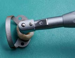
When removing a post, it is critical to break the seal between the post and the tooth structure. This can be accomplished initially through the use of a surgical length 1/4 round bur. This is technique-sensitive, so be careful that you are going along the long axis of the root. Once you have trephined around the post, you can place an ultrasonic tip into the trough. A basic spreader tip will do fine. This will further break the cement or resin and you will soon notice motion in the post. An alternative way to break the seal is to initially use a spreader or troughing tip. Sometimes you can place a spreader tip against the post itself. This works well if the seal has been broken. Don’t rush when removing posts; take your time and don’t panic. The post will come out.
We also strongly recommend using spreader tips with water spray. If you run a spreader tip dry, bad things happen. For one thing, the handpiece on some units becomes very hot and the ultrasonic tip gets overheated. Secondly, the post becomes heated and this heat can eventually be transferred to the periodontal ligament. Lastly, when using an ultrasonic without water spray, you will have an increased smell of burning dentin. Nice, huh? These dilemmas can be avoided through the use of an ultrasonic tip with a water port. If you want to use the ultrasonic “dry” for a few seconds, simply turn off the water. More importantly, the water is there when you need it.
Furthermore, it is obvious that a fiber optic-based ultrasonic will greatly enhance the ability of a clinician to remove a post. Enhanced visual access will ensure a more conservative removal of the post. If too much tooth structure is destroyed during removal of a post, it will complicate the restorative aspect of the tooth and will most likely decrease the overall prognosis.
Removing separated instruments
Sadly, it is a fact of life that some endodontic instruments can break. Fortunately, piezo electric ultrasonics are excellent for removing separated instruments. However, you must be aware of the importance of the location of the separated file. If a rotary file is separated past the canal curvature, this will be extremely difficult to remove. Don’t fall prey to those “experts” who cavalierly say, “I just pull over my microscope and remove it.” These are difficult cases. On the other hand, broken instruments in the coronal half of the canal can be removed in a relatively straightforward manner. However, be honest with yourself - if you do not have the experience, or necessary magnification, you may want to refer this case.
When removing a broken instrument in the coronal third, a thin spreader tip will work nicely. Most ultrasonic companies have tips specifically designed to remove broken instruments. Take the tip into the canal and work it in a counter-clockwise motion around the broken fragment. This will generally dislodge the broken instrument. However, please note that instruments separated in the middle or apical third of a canal are best removed by an experienced clinician with the use of a microscope.
Piezo electric ultrasonics have a host of other indications. For example, poorly fitted crowns can be loosened by placing a vibratory tip against buccal surfaces. Another indication is the condensation of certain restorative materials. However, the most extensive use of these units, besides endodontics, is in hygiene and periodontics; a fiber optic-based ultrasonic makes great sense for any periodontist or hygienist. It is all about enhanced visual access and better treatment for the patient.
We have attempted in this brief article to convey our enthusiasm for the new “Varios 350” ultrasonic unit. It is the combination of both a miniature size and powerful fiber-optic source that truly makes this a next generation unit. It introduces the clinician to an entirely new dimension in ultrasonic use.
Dr. Dennis Brave is a diplomate of the American Board of Endodontics and a member of the College of Diplomates. In endodontic practice for 27 years, he was the senior managing partner of a group specialty practice. Dr. Brave, formerly an associate clinical professor at the University of Pennsylvania, currently holds a staff position at The Johns Hopkins Hospital. Dr. Kenneth Koch is the founder and past director of the program in postdoctoral endodontics at the Harvard School of Dental Medicine. In addition to having maintained a private practice limited to endodontics, he has written numerous articles on endodontics and maintains a faculty position at Harvard. They can be reached at Real World Endo at (866) 793-3636 or through www.realworldendo.com.


