Is it a concussion or TBI? The latest motion-guided imagery and technology can bridge the gap
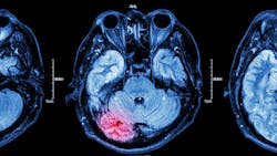
When a patient arrives in your office after mouth or jaw trauma, how can you tell if they had a concussion or traumatic brain injury (TBI)?
According to BMC Oral Health, "traumatic brain injuries are among the most challenging conditions to accurately diagnose in children, and many TBIs are underdiagnosed."1 A 2021 study reported that dental injuries and brain injuries are not independent, and that "it is crucial that dentists who treat traumatic dental injuries rule out concomitant brain injuries."2
You can ask questions, run tests, and take traditional x-rays, but these methods do not provide the data necessary to see what's really going on. These types of scans only provide a snapshot of the spine in a fixed position, so you receive a static image at one point in time. Therefore, it is impossible to see the full scope of what is happening with the spine and brain.
Advantages of real-time motion-guided imaging
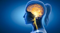
Real-time motion-guided imaging with qEEG and brain-spine communication metrics provide medical evidence of both brain and spinal dysfunction so you can provide a better diagnosis and treatment.
Digital video images are taken to detect vertebral alignment in motion while stabilizing individual vertebral levels rather than offering a single image. This non-invasive assessment of both the brain and the spine provide quantitative, objective, and visual data (down to the tenth of a millimeter) that is consistent, reliable, and accessible via a HIPAA compliant cloud-based system.
When patients also receive a brain baseline assessment, it's possible to track improvements or changes (especially after injuries) of brain performance and spinal motion. An FDA-cleared qEEG with an ERP can be used to take a map of the brain and obtain a benchmark of "normal" brain activity.
The power of modern technology
Brainwave patterns can signal cognitive changes months to years before symptoms emerge. With a set baseline and regular scans, subtle shifts can be detected early. This enables preventive care to help maintain cognitive vitality.
According to Lisa Burris, InControl Imaging cofounder, "Traditional scanning overlooks issues related to communication between the brain and the spine, especially destabilized neck ligaments, which is a common factor in post-concussion syndrome. By using qEEG to pinpoint abnormal brainwave activity from concussion or TBI along with motion guided imagery and brain baseline assessments, doctors and patients receive a complete communication panel of what is being disrupted and where. It's the difference between simply tracking symptoms and actually proving the injury."
Research shows that new technology is making a difference in diagnoses. For example, a study of 196 individuals (119 with chronic neck pain post-whiplash, 77 healthy controls) found that video fluoroscopy detected vertebral instability with 93% sensitivity and 79% specificity. Positive predictive value (PPV) and negative predictive value (NPV) both reached 88%. Cases with four or more abnormal findings returned a PPV of 100%.3
Know what's going on
In the past when patients arrived at your office with chronic jaw pain, bite issues, ear ringing, or facial nerve pain, you were limited to mechanical explanations. Today's new technology allows you to have more clarity. Here are a few of the benefits:
-
It can reveal TMJ dysfunction and whether cervical ligament instability is worsening jaw alignment or bite.
-
It shows whether trigeminal, facial, or cervical nerve irritation is tied to a spinal miscommunication rather than purely dental mechanics.
-
It demonstrates how TMJ or airway issues can alter brain activity, leading to chronic pain, sleep disruption, anxiety, or cognitive fog.
Downsides of traditional scans
Results from traditional X rays, MRIs or CT scans can appear "normal," but patients may continue to suffer with symptoms such as post-concussion syndrome, cognitive dysfunction, headaches, and more. Now, it's possible to know if the root cause of pain is strictly a dental issue or originating from the nervous system
"Traditional X-ray scans are being replaced with the latest technology so that dentists can get real-time data to quickly to treat patients," states Dr. Arth Patel of The Dental Home, https://thedentalhomeofmoore.com. "By combining motion guided imaging with brain scans, it's possible to discover previously missed head injuries, TBIs, concussions and more to get an accurate diagnosis of what's going on with a patient and provide the appropriate treatment in a timely manner."
Recognizing that dental and maxillofacial injury may signal underlying brain injury or concussion is essential. Standard imaging and clinical history are necessary, but they are no longer sufficient to fully assess and manage most dental injury cases. By using the latest guided motion imagery, qEEG, and brain baseline technology, it's possible to detect previously missed issues to assist in treatment, planning, and recovery.
Early intervention is key when it comes to concussions, TBIs, and brain injuries. As medical professionals who are often the first to see a jaw or mouth injury, it's essential to know what's available to provide the best treatment. Not only will this help patients reduce their pain immediately, but it will also assist them in avoiding long-term consequences of untreated brain injuries in the future.
References
- Suprabha, BS, Wilson, ML, Baptist, J, et al. Association of maxillofacial injuries with traumatic brain injuries in paediatric patients: a case-control study. BMC Oral Health. 2024 26;24(1):1560. doi:10.1186/s12903-024-05366-4
- Azadani EN, Townsend J, Peng J, et al. The association between traumatic dental and brain injuries in American children. Dent Traumatol. 2021;37(1):114-122. doi:10.1111/edt.12611
- Freeman MD, Katz EA, Rosa SL, Gatterman BG, et al. Diagnostic accuracy of videofluoroscopy for symptomatic cervical spine injury following whiplash trauma. Int J Environ Res Public Health. 20205;17(5):1693. doi:10.3390/ijerph17051693
About the Author

Melanie Rembrandt
Melanie Rembrandt is an award-winning publicist, author, content strategist, and speaker who helps overwhelmed entrepreneurs and medical professionals have more time to thrive! She specializes in boosting sales, awareness, and credibility, with a unique combination of targeted SEO copywriting, ghostwriting, and public relations. Get more information at Rembrandt Communications®, https://www.rembrandtwrites.com.


