Single-Tooth Implant Treatment in the esthetic zone
Treatment of the anterior maxilla has long proven to be one of the most challenging areas in implant dentistry.1 Managing the esthetics of the soft and hard tissue is just as important as the esthetics of the intended restoration.
New materials such as Procera®, the all-ceramic abutment, have expanded the possibilities for achieving optimal results.2 A wealth of adjunctive therapies also have developed that may enhance the final outcome of single-tooth restorations. Teeth may need to be orthodontically moved. The hard or soft tissue may need to be modified or enhanced after extraction. Decisions also must be made about how and when to provisionalize the implant and place the final restoration. Moreover, every case is different.
The practical consequence is that not every single-tooth case can be treatment-planned the same way. A number of strategies can make treatment planning less daunting, however. Comprehensive initial clinical evaluation — including assessment of the hard and soft tissue, the smile line, the status of the papillae, the occlusion, and other factors — should be done well before the implant is placed. Involvement of the laboratory at the very beginning to discuss the different material options and restoration design is crucial, as is communication with the surgeon. When a team approach is taken and the desired esthetic results are clearly envisioned from the outset of treatment, the chances of maintaining the papillae and harmonizing the soft tissue, emergence profile, and appearance of the restoration will be significantly enhanced.
The following case illustrates how careful planning can help the dentist go beyond offering a limited solution for a problem to ultimately transforming a patient's appearance.
Patient history
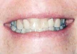
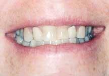
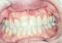
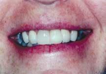
The patient was a 40-year-old female with a moderately high smile line (Figure 1). She presented with a mobile left central incisor that was also discolored and misshapen. Crowns placed years earlier on Teeth Nos. 7 and 10 were also unesthetic, and Teeth Nos. 6, 8, and 11 were malpositioned and discolored as a result of tetracycline therapy that the patient had received as a child. The patient wanted to know the options not only for treating Tooth No. 9, but also for improving her overall smile.
Treatment plan
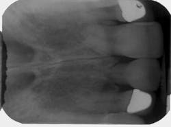
Initial examination of the patient included comprehensive periodontal and restorative examinations and charting; full-mouth radiographs; and evaluation of the smile line, occlusion, hard- and soft-tissue architecture, and interdental/interarch space. The site of the mobile central incisor was found to be free of any pathology. Radiographic examination revealed that the root of Tooth No. 9 was severely resorbed (Figure 2), and the tooth's long-term prognosis was judged to be poor. A decision was made to have the mobile central incisor extracted and immediately replaced with an implant. Immediate placement has been demonstrated to help preserve the hard- and soft-tissue architecture.3,4
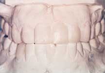
To further improve the patient's appearance, veneers were recommended for the discolored teeth (Nos. 6, 8, and 11), along with replacement of the existing crowns on Teeth Nos. 7 and 10. A diagnostic wax-up helped the patient visualize the esthetic goals, and she agreed to proceed (Figure 3).
Treatment
The surgeon, Dr. Chris Richardson, extracted the tooth, and immediately placed a 13 mm Branemark® externally hexed implant with the osseoconductive TiUnite™ surface.5 A surgical stent was used to optimize placement. The surgeon attached a healing abutment and approximated the tissue over the abutment to promote abundant soft-tissue growth. The patient then proceeded immediately to the prosthodontist's office, where the unesthetic crown on Tooth No. 10 was removed. A new provisional crown was fabricated, and an ovate pontic, intended to replace Tooth No. 9, was cantilevered on Tooth No. 10. The pontic was contoured to conform to the soft tissue and help develop the gingival margin and the papillae adjacent to site No. 9.
The implant healed uneventfully for four months, then it was uncovered. In the restorative office, an immediate, screw-retained provisional restoration was placed. Placing the restoration immediately allows for optimal healing and training of the soft tissue to reestablish the correct gingival margin and papillae. Properly placing the proximal contacts of the restoration less than 5 mm from the osseous crest of the natural tooth promotes papillae formation (Figure 4).5,6
During the subsequent five-month healing period, the patient received an implant in another area of her mouth (Tooth No. 27), and opted to defer final restoration of the anterior maxillary teeth while she recovered from this procedure. Nine months after the initial implant surgery, she returned to the restorative office where the remaining anterior maxillary teeth (Nos. 6, 7, 8, and 11) were provisionalized with a crown and veneers. Retraction cord was placed around all the teeth except for No. 9, where an impression coping was placed. A polyvinylsiloxane impression was made and sent to the dental laboratory, along with photographs, diagrams, and diagnostic casts of the patient's teeth.
Six weeks later, the veneers, the crowns, and a screw-retained porcelain-fused-to-metal final restoration were delivered concurrently. The patient was effusive about her satisfaction with her transformed appearance.
Conclusion
Treatment of a hopeless tooth in the esthetic zone depends substantially on early intervention to help preserve the hard- and soft-tissue architecture and enhance esthetics. Planning of the appearance of the entire dentition, along with coordination among the implant team (surgeon, restorative dentist, and laboratory) are invaluable tools in achieving this end. Implant site development and co-localization of the dental implant with the desired restoration are paramount in achieving optimal esthetics and biomechanical function.
References
- Lewis S. Anterior single-tooth restorations. Int J Periodontics Restor Dent 1995; 15(1):31-41.
- Hegenbarth EA. Procera aluminum oxide ceramics. A new way to achieve stability, precision, and esthetics in all-ceramic restorations. QDT 1996; 1921-34.
- Wohrle PS. Single-tooth replacement in the aesthetic zone with immediate provisionalization: 14 consecutive case reports. Pract Periodont Aesthet Dent 1998; 10(9):1107-1114.
- Petrungaro PS. Immediate restoration of dental implants after tooth removal: a new technique to provide esthetic replacement of the natural tooth system. Int Mag Oral Implantology 2001; 2:6-16.
- Glauser R, Schupbach P, Lundgren AK, Gottlow J, Hammerle CHF. Machined and oxidized microimplants retrieved from humans: a comparison using histomorphometry and microcomputed tomography. Clin Oral Impl Res 2002; 13:4.
- Tarnow DP, Magner AW, Fletcher P. The effect of the distance from the contact point to the crest of bone on the presence or absence of interproximal dental papilla. J Periodontol 1992; 63:995-996.
- Choquet V, et al. Clinical and radiographic evaluation of the papilla level adjacent to single-tooth dental implants. J Periodontol 2001; 72:1364-1371.
Karen McAndrew, DMD, MS
In 2001, Dr. McAndrew founded the Virginia Center for Prosthodontics and Dental Implants in Richmond. She is a clinical assistant professor of oral and maxillofacial surgery at Virginia Commonwealth University School of Dentistry and serves as president of the Virginia section of the American College of Prosthodontists.
