Implants: Immediate Implant Fixture Placement With Immediate Provisional Load for the Edentulous Mandible
a case report
Much has been written on treating the edentulous mandible with clinicians recognizing the edentulous patient as a complex patient population.1 The completely edentulous patient is often rendered frustrated and compromised with the altered function and limitations of complete denture prosthetics. Those with mandibular dentures often suffer the greatest impairment with a majority of those considered as “dental cripples.” It was these patients that Dr. P.I. Branemark originally targeted for the use of dental implants.
Dental implants aid in the retention and stabilization of a prosthesis replacing the completely edentulous arch, while also providing the benefit of long-term bone maintenance. It has been well-documented that dental implants provide a highly predictable, long-term prognosis in the replacement of missing teeth.2,3
The trend in surgical and restorative implant dentistry has been toward immediate placement of the dental fixtures with immediate load of these fixtures. 4,5 Patient desires have pushed the envelope toward early function while minimizing the inconvenience of conventional transitional prostheses during the healing from extraction and implant placement. Immediately placing dental implants at the time of tooth extraction has yielded favorable, predictable results, while early load of these immediately placed dental implants has been studied and has been equally met with predictable results.6 Advantages include better bone and soft-tissue preservation, reduced postoperative pain, significant reduction of clinical chairtime, and greater patient acceptance.
In the edentulous patient, the literature supports immediate placement and immediate load in the mandible using cross-arch stabilization of the implants and a fixed passive-fitting prosthesis on multiple implants having verifiable primary stabilization upon placement.7 Immediate load is only successful if long-term osseointegration accompanies the immediate function. Osseointegration is described as “a direct structural and functional connection between ordered living bone and the surface of a load-carrying implant.”8 Immediate load, in conjunction with osseointegration, is achieved only when micromovement of the dental implant does not exceed the threshold for which osseointegration can occur during the early period of healing of the implant fixture.9,10 This case report provides a simplified approach to the immediate placement and immediate load concept for the completely edentulous mandible.
Case report
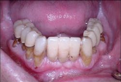
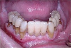
A healthy 55-year-old male presented for examination with a chief complaint being recurrent infection around multiple maxillary and mandibular teeth. Clinical examination revealed generalized severe periodontal disease with hopeless maxillary and mandibular teeth. These teeth were Class II mobile with areas of localized suppuration and failing existing prostheses (Figure 1).
The maxillary arch was treated via full-arch dental extraction with placement of an immediate interim denture. Bilateral sinus augmentations were performed and after adequate healing, eight Nobel Biocare Replace Select implants were placed with the aid of a surgical template.
Procedure
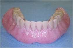
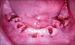
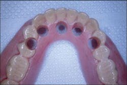
An interim immediate complete mandibular denture was fabricated prior to extraction of the mandibular teeth (Figure 2). The mandibular teeth were extracted and necessary alveoloplasty was performed (Figure 3).
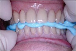
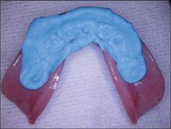
The interim denture was then placed intraorally and a bite registration made (Figure 4) to be utilized in the conversion of this removable prosthesis to the fixed provisional restoration. This registration will be utilized for reapproximation of the denture intraorally once the dental implants and appropriate abutments have been placed.
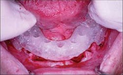
A surgical template was fabricated in clear orthodontic resin by duplicating the mandibular interim denture using a denture duplicating flask. Surgical guide holes were then drilled in the surgical template depicting the exact location of the desired implant placement. Ideal placement of these guide holes is lingual to the anterior teeth and through the occlusal surface of the posterior teeth.
Figure 1 - Pretreatment photograph of mandibular teeth
Figure 2 - Interim denture ready to be converted to fixed provisional restoration
Figure 3 - Mandibular ridges immediately post removal of mandibular teeth
Figure 4 - Bite registration with interim denture in place
Figure 5 - Surgical template placed intraorally to facilitate implant placement within the parameters of the desired prosthesis
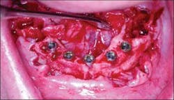
Figure 6 - Immediate placement of Nobel Replace Select fixtures after extraction of mandibular teeth
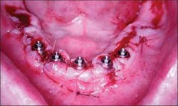
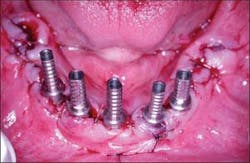
Figure 7 - Multiunit abutments placed and gingiva contoured and sutured
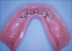
Figure 8 - The antaglio surface of the prosthesis depicting the imprinted surface of the multiunit abutments on the polyvinyl material
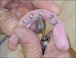
Figure 9 - Preparation of the prosthesis to pick up the temporary cylinders and convert the interim denture into the fixed provisional restoration
Figure 10 - Temporary cylinders placed directly onto the multiunit abutments
Figure 11 - Luting the temporary cylinders to the prosthesis with pink methylmethacrylate
Figure 12 - Prosthesis removed from the mouth with temporary cylinders luted into place
Figure 13 - Trimming and contouring of the prosthesis
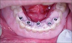
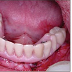
Figure 14 - Immediate delivery of the provisional fixed prosthesis at the time of dental implant placement
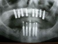
Figure 15 - Panoramic radiograph verifying dental implant placement and immediate load of the mandibular implants with a fixed prosthesis
The surgical template was placed intraorally and utilized in implant placement, co-localizing the desired implant placement with the desired prosthesis (Figure 5). Five 4.3 mm x 13 mm Nobel Biocare Replace Select fixtures were placed in the anterior mandible immediately after extraction of the mandibular teeth (Figure 6). Minor autogenous bone grafting was utilized to repair minor bone defects.
Transmucosal multiunit abutments of appropriate tissue depth were selected and placed onto the implant fixtures and the soft tissue sutured, allowing for the platform of the abutments to be slightly coronal to the gingival margin (Figure 7).
The mandibular prosthesis was aligned with the maxillary prosthesis intraorally using the previously recorded bite registration. Polyvinyl siloxane material was placed on the antaglio surface of the mandibular denture. The patient was guided into centric relation, thus capturing the imprinted location of the multiunit abutments onto the polyvinyl siloxane material (Figure 8). The prosthesis was then removed and holes were drilled corresponding to the location of the abutments (Figure 9).
Temporary cylinders were then placed on the multiunit abutments (Figure 10) and verified that they fit through the holes in the denture. The occlusion was verified and adjusted, and the cylinders were then luted to the prosthesis with cold-cure resin (Figure 11). The temporary cylinders were then unscrewed and the prosthesis was removed from the oral cavity along with the luted cylinders (Figure 12).
In the laboratory, tissue-color cold-cure methylmethacrylate filled in the voids around the temporary cylinders, and the undersurface of the prosthesis was filled in to form a convex, cleansable surface. The prosthesis was trimmed, removing the buccal and lingual flanges and shortening the dentition to first-molar occlusion (Figure 13). The prosthesis was then polished after placing polishing protectors on the undersurface of the temporary cylinders.
Upon delivery of the prosthesis to the patient, the occlusion was verified, the screws hand-tightened, and a cotton pellet and Cavit™ were placed in the access hole (Figure 14). Instructions for care and maintenance were provided to the patient with emphasis on a relatively soft diet for six to eight weeks with no parafunctional loading of the implants (Figure 15). After healing, final impressions will be made and a definitive prosthesis fabricated and delivered.
Conclusion
Fixed immediate provisionalization at the time of implant placement has many benefits for the completely edentulous patient. Transitioning the dentate patient to complete edentulism bears many physical, functional, and psychosocial challenges. Current trends and technology in implant dentistry have significantly reduced the challenges these patients often face. Specifically, immediate implant placement and immediate load with transitional fixed prostheses allow patients to function with minimal transition to the edentulous state. Benefits include minimal swelling and discomfort with little to no functional challenges in conjunction with a decreased healing time.
Through the utilization of a fixed detachable provisional restoration, the dentate patient, using this procedure, never has to experience complete edentulism and the challenge of utilizing a completely removable prosthesis.
Primary stabilization of the dental implant is imperative to facilitating a predictable immediate load. Cross-arch splinted stabilization of the prosthesis on the dental implant complex along with an evenly dispersed occlusal load is necessary for success of this procedure. Many authors have written on the benefits and predictability of immediate placement and immediate load.7 The trend in implant dentistry is toward immediate fixture placement, shorter healing times, and early delivery of the prostheses with benefits for the patient and clinician.
References
1 Zarb GA, Schmitt A. Osseointegration and the edentulous predicament. The 10-year-old Toronto study. Brit Dent J 1991; 170(12):439-44.
2 Adell R, Eriksson B, Lekholm U, Branemark PI, Jemt T. A long-term follow-up study of osseointegrated implants in the treatment of the totally edentulous jaws. Int J Oral Maxillofac Implants 1990; 5:347-359.
3 Preiskel HW, Tsolka P. Treatment outcomes in implant therapy: the influence of surgical and prosthetic experience. Int J Prosthet 1995; 8:3:273-279.
4 Balshi TJ, Wolfinger GJ. Teeth in a day. Implant Dent 2001; 10:231-233.
5 Malo P, Rangert B, Nobre M. All-on-four: immediate-function concept with Branemark system implants for completely edentulous mandibles: A retrospective clinical study. Clinical Impl Dent 2003; 5:(1)2-9.
6 Nikellis I, Levi A, Nicolopoulos C. Immediate loading of 190 endosseous dental implants: A prospective study of 40 patient treatments with up to two-year data. Int J Oral Maxillofac Implants 2004; 19(1):116-123.
7 Chiapasco M. Early and immediate restoration and loading of implants in completely edentulous patients. Int J Oral Maxillofac Implants 2004; 19 (suppl):76-91.
8 Branemark PI, Zarb G, Albrektsson T, eds. Tissue integrated prostheses. Osseointegration in Clinical Dentistry. Chicago Quintessence Publ Co Inc; 1985.
9 Schnitman PA, Wohrle PS, Rubentain JE Immediate fixed interim prostheses supported by two-stage threaded implants: methodology and results. J Oral Implantology 1990; 16:96-105.
10 Schnitman PA, Wohrle PS, Rubentain JE, DaSilva JD, Wang NH. Ten-year results for Branemark implants immediately loaded with fixed prostheses at implant placement. Int J Oral Maxillofacial Implants 1997; 12:495-503.
Karen McAndrew, DMD, MS
Dr. McAndrew maintains a private practice in Richmond, Va., limited to prosthodontics at the Virginia Center for Prosthodontics and Dental Implants. She holds an assistant professor appointment in the Department of Oral and Maxillofacial Surgery at the VCU School of Dentistry.
Kanyon R. Keeney, DDS,
Dr. Keeney maintains a private practice in Richmond, Va., limited to oral and maxillofacial surgery focusing on dental implant therapy. He holds an assistant professor appointment in the Department of Oral and Maxillofacial Surgery at the VCU School of Dentistry and is a diplomate of the International Congress of Oral Implantology.
