The extraoral and intraoral dental exam process for complete patient health
Dentists have the responsibility to evaluate, diagnose, and treat not only the dentition but the intraoral and extraoral structures and tissues as well as the overall health of our patients. When it comes to the intra-, extra-, and perioral tissues, our awareness of normal, abnormal, and variant from normal and how we define these terms are critical to diagnosis and management. Looking beyond the dentition and periodontium is an important step in overall patient assessment. None of us would want to focus on a patient’s crowns for the maxillary anterior teeth while a lesion of significance goes unnoticed in another region of the oral cavity. These undiscovered or overlooked problems are issues not only from a medicolegal aspect but our own oath to provide the best and most comprehensive health care possible. As dental providers, we must take time to be thorough, comprehensive, and diligent during the examination process, management of the issue(s), and requisite follow-up.
Our patients often present with a particular concern, and we (and our auxiliary staff) focus on just that—the chief complaint. We even have a code to use for this: ADA Code D0140 limited oral evaluation – problem focused. But we should never limit ourselves to just “the problem.” We need to have an adequate medical history to treat, and we need to assess more than just the immediate intraoral area where the problem manifests. A complete intraoral and extraoral surveillance will allow for a complete understanding of the patient’s oral status and overall health. If something else is present, pathological, and hiding in plain sight, “problem focused” won’t give us a pass in the courtroom or in our conscience. Do the right thing for yourself and your patient: evaluate, document, and manage.
Related reading:
- The state of oral and oropharyngeal cancer screening in our profession
- Clinical lifesavers: Security in your oral cancer screening technology
I will share some of my personal preferences for evaluating and managing patients. These techniques work well in my hands and with my patients. Like with many things in dentistry, we all have our own methods. The key is the result, which is an appropriately examined and managed patient, so that when all is said and done, we have confidence that we did everything in our power and diagnostic ability to ensure that no potential problems exist. We can never be 100% certain, but with certainty we will know we were thorough.
General assessment and overview
My process commences with a general assessment and overview for symmetry and extraoral evaluation for suspicious lesions in the head and neck region. These can be difficult conversations because the patient is usually aware of such lesions and may feel you should not extend your view beyond the oral cavity. However, many patients have not been evaluated by a physician and don’t know that we have knowledge, experience, and understanding of when a lesion should be evaluated by a physician or dermatologist. Patients may say, “That’s been there forever” or “It’s nothing,” but it is our responsibility to recommend a further evaluation.
Extraoral exam
I then advise the patient that I will perform a thorough extraoral exam. I say I am evaluating the head and neck to make sure everything appears healthy; I’ll say I am assessing for any abnormalities. I do not tell them I am “looking for cancer” or “performing an oral cancer screening.” I have heard students and practitioners state to patients, “I am now going to check your mouth for cancer.” The moments during your inspection and evaluation will be anxiety-ridden for the patient. Any positive findings for reevaluation or follow-up will instill fear and possibly be a source of unnecessary stress until the patient is cleared or diagnosed though appropriate testing.
Telling a patient that they are “cancer free” after your evaluation is never infallible. This could lead to a false sense of security. A confirmation is not possible with certainty for any lesion or growth without further testing, imaging, and/or biopsy to confirm a diagnosis of cancer. I use the terminology “oral cancer screening” in my recorded notes, but I do not want to verbally imply to a patient that I am capable of diagnosing cancer through my visual inspection or palpation. I need my patients to act responsibly when there is concern, not be obsessed with worry, and never let their guard down by ignoring suspicious changes. A diagnosis of dysplasia or precancerous cellular changes requires further follow-up and evaluation if changes are noted. Continued surveillance, even without a cancer diagnosis, is a must for areas with suspicious tissue changes.
I start my process by palpating the lymph nodes of the head and neck: preauricular, postauricular, occipital, submandibular, submental, cervical nodes following the anatomy of the neck divided into anterior and posterior triangles demarcated by the midline, clavicle, and sternocleidomastoid. This will also involve the midline structures of the neck, including an evaluation of the thyroid for symmetry and enlargement. Modifying head position—left, right, chin up, chin down—is important to palpating those abnormalities that attempt to remain hidden. If abnormalities were obvious and visually evident, our jobs would be much easier. Any positive findings of lymphadenopathy, tenderness, lack of symmetry, or enlargement require follow-up and appropriate referral when a reasonable diagnosis or etiology cannot be established.
Intraoral exam
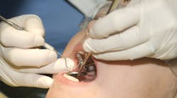
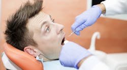
After a general overview and complete head and neck exam, I progress to the oral cavity from the outside to the inside. Start with a visual inspection and palpation of the labial and buccal hard tissues of the gingiva and palate—the tongue, lateral borders, dorsum, and ventral surface—using gauze and a mirror to visualize all surfaces. Use of indirect vision with a mirror and excellent lighting is essential. Magnification is very helpful, and alternate light sources can also expose variations that are not visible under natural or operatory lights.
Next, I move to the floor of mouth, using bimanual and bidigital palpation, meaning one hand in the submandibular area and one intraorally so I can palpate any displaced abnormalities between opposing digits. Gentle pressure should be enough to identify the normal anatomy or abnormalities but should not be painful or cause unnecessary discomfort to the patient. Localized pain can be a sign of a problem, requiring further evaluation, follow-up, or referral.
As we proceed further to the posterior, we should visualize the oropharynx. The ability to view these structures is variable between patients. To the best of our ability and based on the patient’s level of compliance, we hope to be able to clearly visualize the right and left anterior and posterior tonsillar pillars and the intervening tonsillar crypt. We also would like to be able to view the posterior pharyngeal wall. Again, note lack of symmetry, tissue changes, ulcerations, enlargement, or anything that is different from what your knowledge of normal is.
As we evaluate the extraoral and intraoral structures, we should be cognizant of the relationship between these areas and the 12 cranial nerves. Note abnormalities of pain, paralysis, lack of function (motor or sensory), all of which could be signs of potential problems. These abnormalities of function and sensation can be indicative of neuropathology or even a cerebrovascular accident or strokes.
Our ability to recognize these deficits in a timely fashion can be lifesaving and should be taken very seriously. We must clearly emphasize our concern and level of urgency to the patient, and action must be immediate. Our ability to separate a medical emergency from a medical concern requiring follow-up is a significant and serious responsibility—an area where we never want to be wrong or fail to act appropriately.
Takeaway
My take-home message is to be thorough, comprehensive, and skilled to knowledgably do your best as well as do the right thing for your patient’s overall health and your peace of mind, knowing you are a responsible and conscientious dental practitioner.
Editor’s note: This article first appeared in Through the Loupes newsletter, a publication of the Endeavor Business Media Dental Group. Read more articles and subscribe to Through the Loupes.


