Pathology case: Extra! Extra! Read all about it!
Case presentation and exam
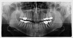
A healthy 45-year-old female presents for her recare exam and an update on her panoramic radiograph. She reports no concerns or complaints.
An assessment of the pan revealed two radiopaque lesions apical to the root tips of nos. 8 and 9, each measuring approximately 8 mm x 4 mm (figure 1). There was no tenderness on palpation in the generalized area. Access to a previous pan taken six years prior revealed no significant changes to the radiopacities (figure 2).

Diagnosis and discussion
Given the location, absence of change, and shape of the lesions, the diagnosis for these lesions is mesiodens, a “supernumerary tooth located between the maxillary central incisors.”1 The lesions may occur “as single, multiple, unilateral, or bilateral (lesions); the presence of multiple supernumerary teeth is called ‘mesiodentes.’”2 By way of appearance, they typically manifest as a conical or peg-shaped form.2
In general, supernumerary teeth—also referred to as hyperdontia, or extra teeth—can occur in any location, but they are more prevalent in the maxilla (90%) as opposed to the mandible (10%).1 The most common type of supernumerary teeth are mesiodentes.2 Their etiology remains unclear, although it is theorized that they are either an isolated finding or part of a “syndrome, specifically cleft lip and palate, cleidocranial dysostosis, and Gardner’s syndrome.”2
Diagnosis is primarily via radiograph and can confirmed by taking multiple angles using intraoral and extraoral radiographs (i.e., panoramic, periapicals, etc.). Furthermore, three-dimensional CBCT scans allow dialing in on the palatal location of the mesiodentes and relationship to adjacent teeth. Delayed or altered eruptions of adult dentition also give rise to the suspicion of mesiodentes. Other common signs include cyst formation and crowding.
Treatment
Treatment of mesiodens varies. Although typically asymptomatic, they are “often extracted for aesthetic reasons, to allow the eruption of other teeth, orthodontic reasons, and/or suspected pathology.”3 In this particular case, the mesiodentes had been stable through the years without pathology or issues with regard to the patient’s permanent dentition. We will continue to monitor the patient via regular panoramic x-rays.
References
- Sapp JP, Eversole LR, Wysocki GP. Contemporary Oral and Maxillofacial Pathology. St. Louis, MO: Mosby; 1997:4-5.
- Meighani G, Pakdaman A. Diagnosis and management of supernumerary (mesiodens): a review of the literature. J Dent (Tehran). 2010;7(1):41-49.
- Hyperdontia. Wikipedia. Updated May 5, 2020. Accessed March 15, 2020. https://en.wikipedia.org/wiki/Hyperdontia.
About the Author

Stacey L. Gividen, DDS
Stacey L. Gividen, DDS, a graduate of Marquette University School of Dentistry, is in private practice in Montana. She is a guest lecturer at the University of Montana in the Anatomy and Physiology Department. Dr. Gividen has contributed to DentistryIQ, Perio-Implant Advisory, and Dental Economics. You may contact her at [email protected].