Pathology case: An almost-vague radiodense lesion
A 50-year-old female presented for a routine new-patient exam. She was on a prescription of Keflex for an infection. The patient was otherwise healthy, taking no other medications and having no medical concerns.
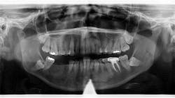
A panoramic radiograph revealed an almost-vague radiodense lesion on the lower left side, around the apices of teeth nos. 20–22, measuring approximately 3 cm x 1.5 cm. The lesion was nonpalpable, the area was not tender to palpation, and the patient was unaware of its presence.
Diagnosis
Differentials
- Idiopathic osteosclerosis
- Sclerosing osteitis
- Periapical cemento-osseous dysplasia (PCOD)
- Focal cemento-osseous dysplasia (FCOD)
Definitive diagnosis: Idiopathic osteosclerosis
The two first differentials are actually related to one another. “An area of bony sclerosis is termed sclerosing or condensing osteitis if its cause can be associated with an inflammatory process and idiopathic osteosclerosis if its cause cannot be readily explained.”1
Discussion and treatment recommendations
The following characteristics are similar between the two lesions.1
- They are almost invariably painless and do not produce expansion of the cortex.
- The covering mucosa is normal in appearance.
- Sinuses are not present, and regional lymph nodes are characteristically asymptomatic.
- Approximately 85% of the sclerosing and sclerosed areas occur in the mandible of whites, where the first molar region is the predominant site; in blacks, approximately 71.6% of focal bony sclerotic areas are in the mandible.
- These sclerotic areas may remain unchanged for years (even after successful treatment of associated infected teeth), may partially resolve, or may completely disappear (and regions become radiographically normal again). Serial radiographs showing any increases in size indicate an active lesion.
Also by Stacey L. Gividen, DDS:
Can and should you use diamond burs more than once?
I’m a "heartless, judgmental, inconsiderate dentist."
Sclerotic lesions can vary in density, shape, and size (mm to cm). Diagnosis includes testing of the involved teeth (pulp testing, etc.) and acquiring any previous radiographs. If there is pathology involved, then the appropriate treatment is recommended for the tooth and possible removal of the lesion. If all tests are within normal limits, it is recommended that subsequent radiographs be taken to note any changes that may take place (e.g., size of lesion, any root resorption, expansion, etc.).
Since this patient presented with no neurosensory changes, cortical expansion or displacement or resorption of adjacent teeth, and its cause could not be readily explained, it is consistent with a diagnosis of idiopathic osteosclerosis. The patient continues to be recalled periodically to assess for changes in this area.
Originally posted in 2021 and updated regularly
Editor’s note: This article appeared in Through the Loupes newsletter, a publication of the Endeavor Business Media Dental Group. Read more articles and subscribe to Through the Loupes.
Reference
- Wood NK, Goaz PW. Differential Diagnosis of Oral and Maxillofacial Lesions. 5th ed. Mosby Publishing; 1997:458–463.
About the Author
Stacey L. Gividen, DDS
Stacey L. Gividen, DDS, a graduate of Marquette University School of Dentistry, is in private practice in Montana. She is a guest lecturer at the University of Montana in the Anatomy and Physiology Department. Dr. Gividen has contributed to DentistryIQ, Perio-Implant Advisory, and Dental Economics. You may contact her at [email protected].

