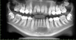
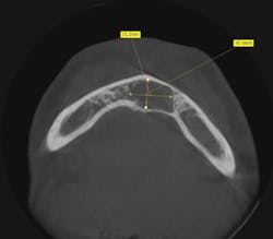
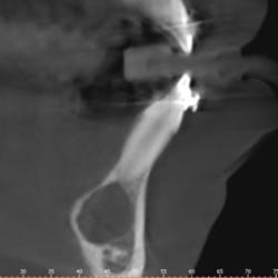
Dr. Ahmad Chaudhry presents the oral pathology case of a 14-year-old male with a lesion on the anterior mandible first seen on periapical x-rays his general dentist took. A CT scan revealed a circular, hypodense area.
Editor's note: This article originally appeared in Breakthrough Clinical, a clinical specialties newsletter from Dental Economics and DentistryIQ. To subscribe, visit dentistryiq.com/subscribe.
Presentation
A 14-year-old male presented with a lesion of the anterior mandible first seen on periapical x-rays taken by his general dentist. The patient did not have symptoms and was unaware of any lesion prior to the x-rays.
Medical history and clinical exam
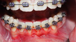
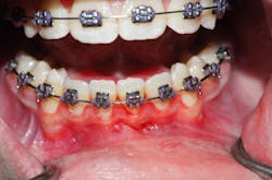
Clinically, there was no buccal or lingual expansion (figure 1). The patient’s medical history was noncontributory. A CT scan revealed a 1.5 cm x 1 cm well-defined, circular, hypodense area inferior to the lower incisors (figures 2–4).
Figure 1
Figure 2
Figure 3
Figure 4
After examining this information, what would be the next step in this case?
Send your answers to [email protected]. Next month, we will present the final diagnosis and recommended treatment for this oral pathology case.
CALL FOR PATHOLOGY CASES
Do you have an interesting oral pathology case you would like to share with Breakthrough’s readers? If so, submit a clinical radiograph or high-resolution photograph, a patient history, diagnosis, and treatment rendered to [email protected].
Editor's note: This article originally appeared in Breakthrough Clinical, a clinical specialties newsletter from Dental Economics and DentistryIQ. To subscribe, visit dentistryiq.com/subscribe.
